A new method for neuroimaging analysis is shown to work with small groups of participants, opening the door for many studies that don’t have access to massive sets of brain images.
Tag: MRI
MD Anderson Research Highlights for June 5, 2024
The University of Texas MD Anderson Cancer Center’s Research Highlights showcases the latest breakthroughs in cancer care, research and prevention. These advances are made possible through seamless collaboration between MD Anderson’s world-leading clinicians and scientists, bringing discoveries from the lab to the clinic and back.
Researchers find key differences in brain development between autistic boys and girls
A new study by UC Davis researchers finds key differences in the development of the cortex between autistic boys and girls ages 2-13.
UC San Diego Receives $6.7M to Develop Whole-Body Inflammation Imaging
Researchers at UC San Diego have received new grants that will help develop full-body imaging techniques to detect inflammation, which is currently very difficult to visualize in the clinic.
A flicker of truth: Piercing the “continuity illusion”
A study by a team at the Champalimaud Foundation (CF) has cast a new light on the superior colliculus (SC), a deep-seated brain structure often overshadowed by its more prominent cortical neighbor.

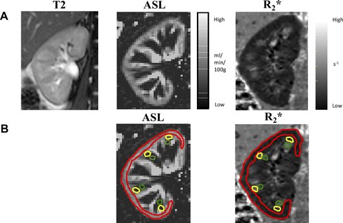
A Noninvasive Way to Measure Placental Health
A healthy placenta is critical for a healthy baby. But unfortunately, there’s no direct way to measure how well this important organ is working.
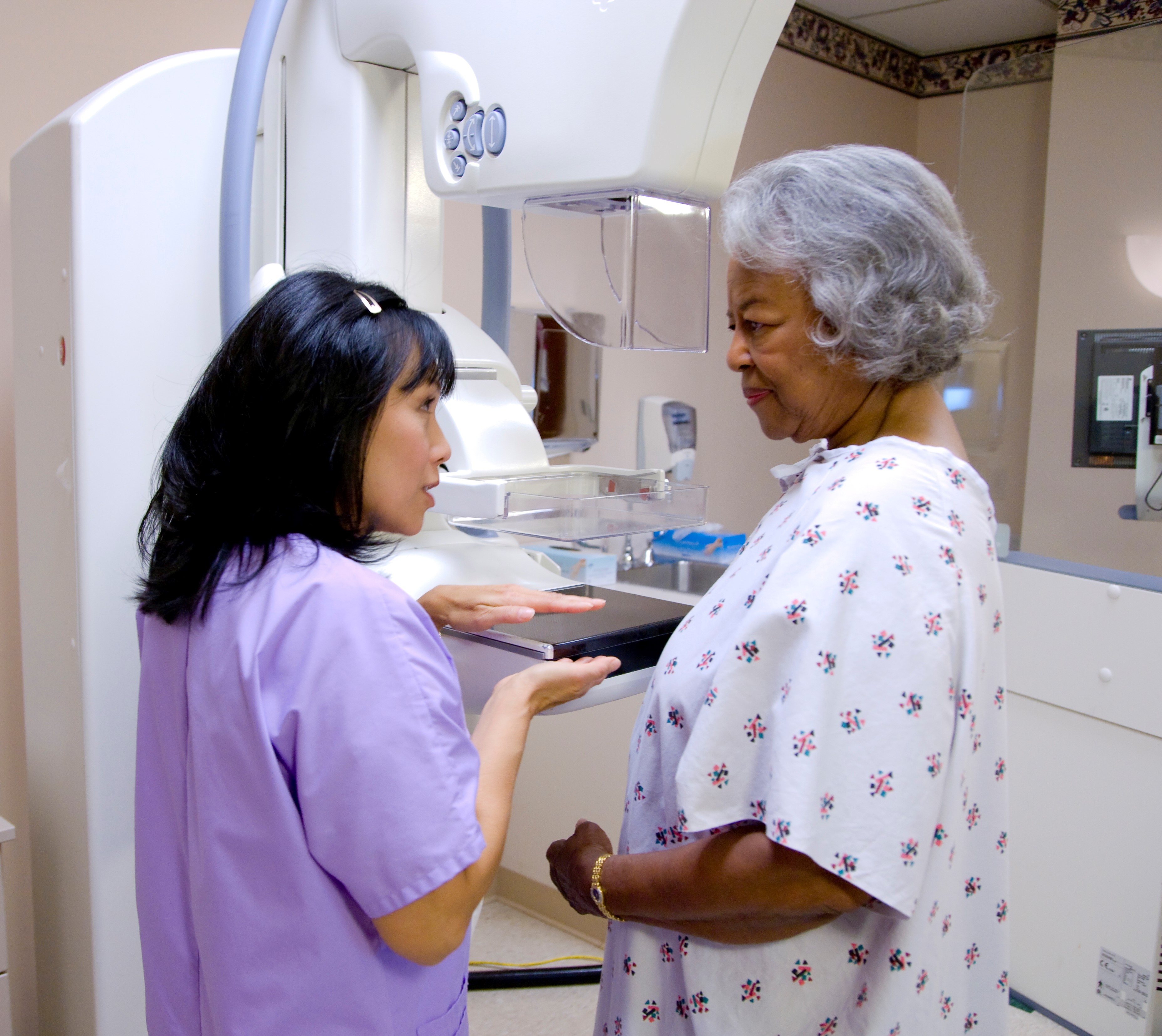
With Regular Screening, More Women Survive Breast Cancer
To mark Breast Cancer Awareness Month, the American Cancer Society highlights its guidelines encouraging average-risk women to begin regular screening mammograms at age 45, with the option to begin screening as early as age 40.
Diabetes linked to functional and structural brain changes through MRI
A new study finds that the longer a person has type 2 diabetes, the more likely they may be to experience changes in brain structure. MRI results, researchers say, indicate the negative effects longstanding diabetes may have on brain health outcomes and emphasize the importance of preventing early onset type 2 diabetes.
Mount Sinai Brooklyn Expands Cancer Services, Infusion Center in $4 Million Project
The Mount Sinai Health System today held a grand opening of the expansion of the Mount Sinai Brooklyn Ambulatory Infusion Center, a cancer treatment center that brings innovative cancer therapy and clinical trials to residents of southern Brooklyn.
Studying brain activity of swallowing helps researchers understand aging, disease
Researchers at the Beckman Institute for Advanced Science and Technology, Carle Hospital, and Purdue University teamed up to develop a new imaging tool that will improve our understanding of how the brain controls swallowing in both healthy patients and those experiencing a swallowing-related disorder. Their work will be funded by a five-year grant expected to total $2.8 million from the National Institute on Aging of the National Institutes of Health.
Study: Drug May Delay Earliest Symptoms of Multiple Sclerosis
A drug called teriflunomide may delay first symptoms for people whose magnetic resonance imaging (MRI) scans show signs of multiple sclerosis (MS) even though they do not yet have symptoms of the disease. The preliminary study, released April 19, 2023, will be presented at the American Academy of Neurology’s 75th Annual Meeting, being held in person in Boston and live online from April 22-27, 2023. Called radiologically isolated syndrome, the condition is diagnosed in people who do not have MS symptoms but who have abnormalities in the brain or spinal cord called lesions, similar to those seen in MS.
Penn Medicine Study Reveals New Insights on Brain Development Sequence Through Adolescence
Brain development follows a newly identified, non-uniform developmental sequence rendering youth to environmental impacts through adolescence.
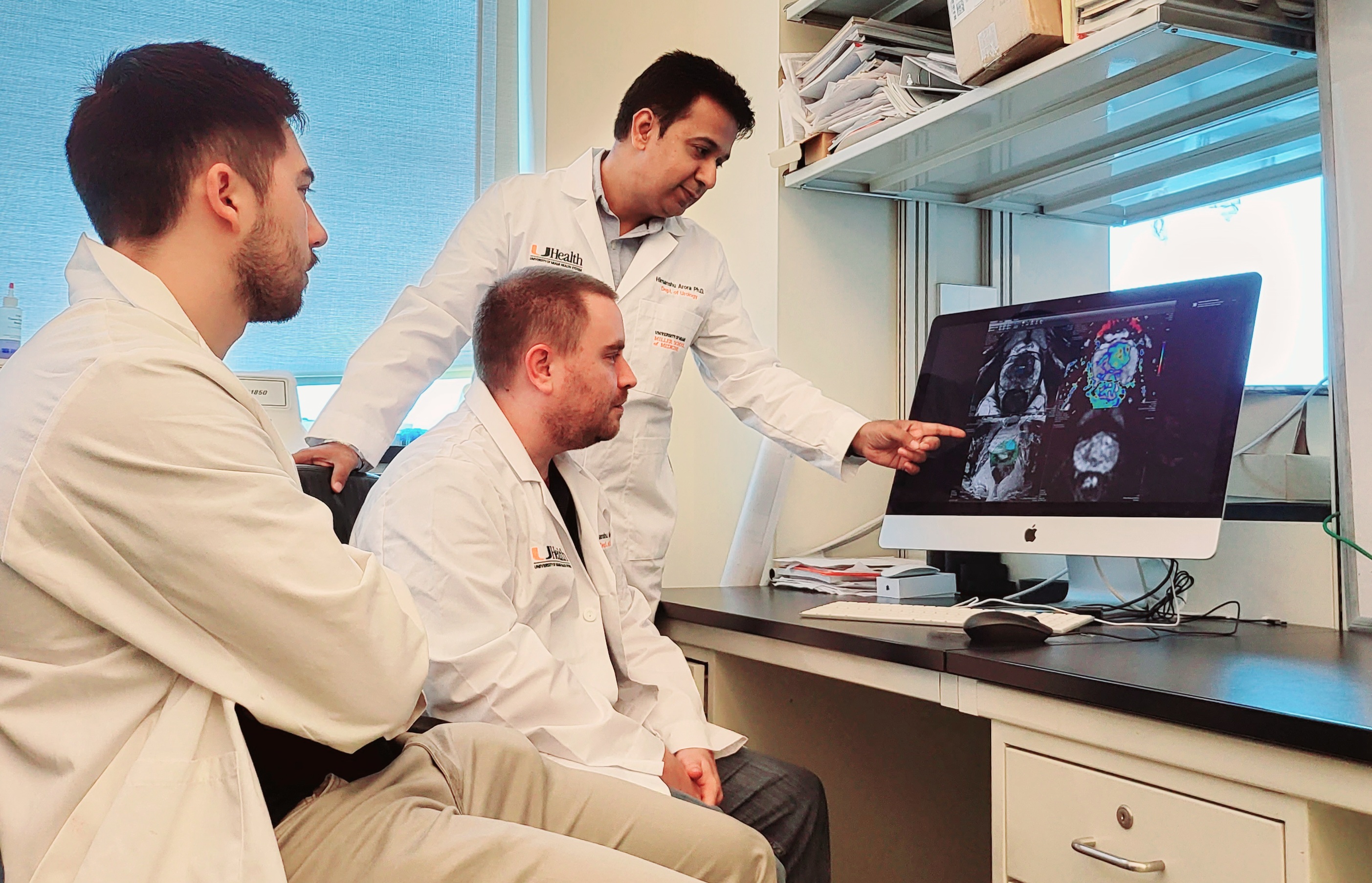
Scientists Pioneer Research to Harness Power of Machine Learning in Prostate Cancer Imaging
Researchers at Sylvester Comprehensive Cancer Center and the Desai Sethi Urology Institute are pioneering research to harness machine learning for the diagnosis and prognosis of prostate cancer, using magnetic resonance imaging (MRI).
MRI turns 50: Expert Brad Sutton explains its history and role in understanding the aging brain
March 16, 2023, marks 50 years since Paul Lauterbur published his seminal Nature paper establishing zeugmatography — now familiar to most as magnetic resonance imaging or simply MRI — as a viable way to visualize objects with a magnetic field…
Case Western Reserve University awarded $3M grant to advance MRI scan and software to analyze aggressive brain tumors more effectively
With a new five-year, $3.03 million grant from the National Cancer Institute—an agency of the National Institutes of Health—Case Western Reserve University researchers are leading the development and commercialization of a novel MRI and software technology that results in more accurate, consistent brain tumor diagnosis.
ICD detection and treatment of arrhythmias not impacted by MRI
A cohort study of more than 600 persons with non-magnetic resonance imaging (MRI)-conditional implantable cardioverter defibrillators (ICDs) found that these ICDs still appropriately treated detected tachyarrhythmias after MRI. The findings are published in Annals of Internal Medicine.
MRI-guided radiotherapy produces fewer side effects and better quality of life for patients with localized prostate cancer
For men who undergo radiotherapy for localized prostate cancer, the precise targeting capabilities of MRI guidance resulted in fewer toxicities and better quality of life according to new research from UCLA Jonsson Comprehensive Cancer Center.
Study Shows Promising Safety, Patient Outcomes Data for MRI-Guided Adaptive Radiation Therapy to Treat Pancreatic Cancer
Findings from a recent prospective study show promising safety and patient outcomes data for locally advanced and borderline resectable pancreatic cancer treatment using ablative Stereotactic MRI-Guided On-table Adaptive Radiation Therapy, also known as SMART.
2D and 3D MRIs provide reliable measurements for planning ACL surgery, UTSW study shows
Magnetic resonance imaging (MRI) can reliably establish measurements for anterior cruciate ligament (ACL) “footprints” that are critical to the placement of grafts for reconstruction surgery
Neuroimaging Study Reveals Functional and Structural Brain Abnormalities in People with Post-Treatment Lyme Disease
In a study using specialized imaging techniques, Johns Hopkins Medicine researchers report distinctive changes in the “white matter” and other brain tissue physiology of those with post-treatment Lyme disease, a condition affecting 10% to 20% of the nearly half a million Americans who contract Lyme disease annually.
Functional, nonepileptic seizures show structural abnormalities in brain scans, study shows
For a long time, functional, nonepileptic seizures were not believed to involve structural changes in the brain, but a new study suggests that they are associated with structural changes that can be seen using MRI. Researchers say the findings bring potential for earlier diagnosis of functional seizures, which are often misdiagnosed as epilepsy.
Can Obesity and Stress Influence Appetite? New Johns Hopkins Study Shows It’s All In Your Head
In a series of experiments using functional magnetic resonance imaging (fMRI) to measure brain activity across networks in the brain, Johns Hopkins Medicine researchers looked at how stress might increase appetite in obese and lean adults.
Ceramic material could improve MRIs by enabling faster times, better images
An academic/enterprise partnership that includes Penn State researchers is developing a new dielectric material to enable magnetic resonance imaging (MRI) machines with shorter scan times and higher image resolutions, good news for cutting the cost of MRI scans for the hospitals and for patients who struggle with MRI-related anxiety.
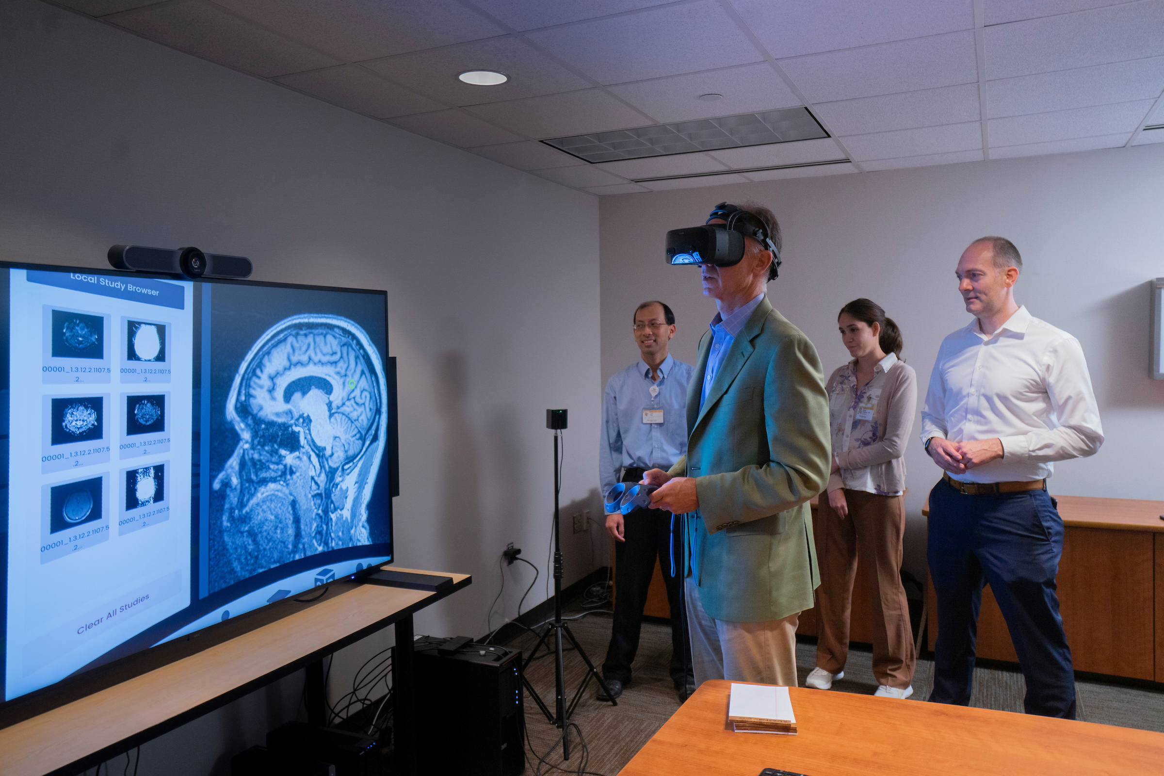
Wake Forest University School of Medicine Collaborating with Physicians in Ecuador on Virtual Reality Radiology Project
Neuroradiologists at Wake Forest University School of Medicine have begun a pilot study testing the use of virtual reality (VR) systems to remotely read MRIs and other medical images.
“The wonderful thing about this is that it’s a collaborative environment with people who can look at medical images at the same time, regardless of where they are in the world,” Burdette said. “We are able to interpret images in incredible detail while communicating with the referring clinicians who are seeing the same images in real time, which is just not possible in a traditional reading room in the hospital.”
Few patients undergo recommended MRI screening after silicone implant breast surgery
Only six percent of women with silicone breast implants followed the previous US Food and Drug Administration (FDA) recommendation for regular magnetic resonance imaging (MRI) screening, suggests a study in the August issue of Plastic and Reconstructive Surgery®, the official medical journal of the American Society of Plastic Surgeons (ASPS).
MD Anderson Research Highlights for July 27, 2022
Clinical advances include treating hematologic cancers with effective targeted therapies, circulating tumor DNA as a biomarker for recurrence with colorectal liver metastases, and using magnetic resonance imaging (MRI) to guide surgical decisions for patients with lateral pelvic lymph node metastases in rectal cancer. Laboratory findings offer new understanding of the pancreatic cancer immune microenvironment, melanoma cell states, TP53 mutation status in acute myeloid leukemia (AML), and potential targets for metastatic prostate cancer and GNAS-mutant colorectal cancer.
Researchers Reveal Brain Changes, Differences in Children with ADHD
UNC scientists conducted a study to image the neural activity analogues to cognitive flexibility and discover differences in the brain activity of children with ADHD and those without.
How MRI and CT predict flap failure after head and neck reconstructive cancer surgery
A new study finds that early postoperative CT scans and MRIs can help predict whether a free flap, used for reconstructive head and neck cancer surgery, will fail. The method carries around a 10-40% risk of wound complications, and researchers say the findings could allow surgeons to intervene earlier if the flap fails.
Visual System Brain Development Implicated in Infants who Develop Autism
For the first time, scientists have found that brain differences in the visual brain systems of infants who later are diagnosed with autism are associated with inherited genetic factors.
Fast-tracked: First in-human trial for aggressive brain tumours
A novel technology designed to precisely image aggressive brain cancers and guide treatment is being developed by the University of South Australia and Australian cancer diagnostic company, Ferronova, potentially helping thousands of people who are diagnosed with the deadly condition each year.
MD Anderson and Siemens Healthineers collaborating to enable consistent clinical implementation of quantitative MRI
MD Anderson and Siemens Healthineers have announced the collaborative development of an education program focused on enabling the implementation of consistent, high-quality MRI in radiation oncology.
New machine learning method to analyze complex scientific data of proteins
Scientists have developed a method using machine learning to better analyze data from a powerful scientific tool: nuclear magnetic resonance (NMR). One way NMR data can be used is to understand proteins and chemical reactions in the human body. NMR is closely related to magnetic resonance imaging (MRI) for medical diagnosis.
Focused Ultrasound Breakthroughs from the Summer of 2021
Here are eight amazing developments in the use of Focused Ultrasound from just the last three months, including: treating cancerous tumours, triggering the targeted release of medicine in the body, immunotherapy, and pain management. See more in the Focused Ultrasound Channel
Portable MRI provides life-saving information to doctors treating strokes
When patients exhibit stroke symptoms, doctors must quickly make a life or death determination: Are their symptoms caused by a clot that can be treated with blood thinners or by bleeding in the brain, which may require surgery?
8 weeks of meditation studies can make your brain quicker
Just eight weeks of meditation studies can make your brain quicker, according to new research from Binghamton University, State University of New York.
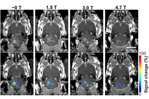
MRI’s magnetic field affects focused ultrasound technology
A new finding prompts researchers, clinicians to consider this impact in future research and clinical treatment of brain diseases
New Research on MRI Biomarkers May Aid in Evaluating Potential Treatments for Peripheral Nerve Diseases
Jun Li, M.D., Ph.D., professor and chair of the Department of Neurology at the Wayne State University School of Medicine, recently received a $246,172 R-21 grant from the National Center for Advancing Translational Sciences of the National Institutes of Health (NIH) to develop new strategies for assessing appropriate treatments of peripheral nerve diseases.
Mouse brain imaged from the microscopic to the macroscopic level
Researchers at the University of Chicago and the U.S. Department of Energy’s (DOE) Argonne National Laboratory have leveraged existing advanced X-ray microscopy techniques to bridge the gap between MRI (magnetic resonance imaging) and electron microscopy imaging, providing a viable pipeline for multiscale whole brain imaging within the same brain
NIH-funded study shows children recycle brain regions when acquiring new skills
Scientists studied the brain activity of school-aged children during development and found that regions that activated upon seeing limbs (hands, legs, etc.) subsequently activated upon seeing faces or words when the children grew older. The research, by scientists at Stanford University, Palo Alto, California, reveals new insights about vision development in the brain and could help inform prevention and treatment strategies for learning disorders. The study was funded by the National Eye Institute and is published in Nature Human Behaviour.
Are Heavy Metals Toxic? Scientists Find Surprising New Clues in Yeast
Scientists at Berkeley Lab and UC Berkeley have compiled the most complete library yet of lanthanide heavy metals and their potential toxicity – by exposing baker’s yeast to lanthanides. Their findings could help researchers uncover hidden pathways between lanthanide metals and disease.
Researchers launch ‘next generation’ human brain imaging lab
Researchers to measure the brain’s subtle magnetic signals in two research volunteers simultaneously as they interact, capturing the rich complexity of the brain’s signaling during face-to-face social interactions in real-time.
Researchers launch ‘next generation’ human brain imaging lab
Researchers to measure the brain’s subtle magnetic signals in two research volunteers simultaneously as they interact, capturing the rich complexity of the brain’s signaling during face-to-face social interactions in real-time.
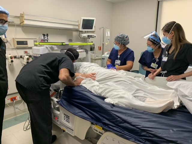
JFK University Medical Center First Hospital in New Jersey to Utilize New Portable MRI
Hackensack Meridian JFK University Medical Center is the first hospital in New Jersey to operate a new portable MRI that can be wheeled to the bedside of critically ill patients. The world’s first portable MRI called Swoop™, enables clinicians to obtain neurological images of critically ill patients at the point of care quickly and conveniently.
LHMC Radiology team reports on model algorithm to optimize MRI scheduling
The first known study exploring optimal outpatient exam scheduling through a model algorithm was shown to yield shorter wait times for magnetic resonance imaging (MRI) for patients and reduced costs.
Researchers Help Pioneer New Era in Prostate Cancer Active Surveillance
Sylvester Comprehensive Cancer Center researchers are looking at ways to combine imaging and biomarkers to predict prostate cancer progression more accurately.
MRI frequently underestimates tumor size in prostate cancer
Improving imaging processes will lead to more successful treatments and help reduce morbidity in men with the disease.
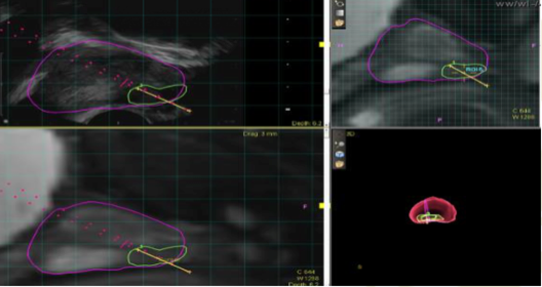
Noninvasive Imaging Effectively Shows Variations in Renal Blood Flow
Renal blood flow changes throughout the day in tandem with the body’s circadian clock, with increasing flow during daytime hours and decreasing flow in the evening and into the night.

¬Children With Asymptomatic Brain Bleeds As Newborns Show Normal Brain Development At Age 2
A study by UNC School of Medicine researchers finds that neurodevelopmental scores and gray matter volumes at age two years did not differ between children who had MRI-confirmed asymptomatic subdural hemorrhages when they were neonates, compared to children with no history of subdural hemorrhage.
‘Fast’ MRI Detects Breast Cancers that 3-D Mammograms May Miss
In a retrospective study of Penn Medicine patients, all of whom had a negative 3-D mammogram within the previous 11 months, abbreviated MRI detected roughly 27 cancers per 1,000 women screened. By comparison, 3-D mammography detects about four to five cancers in 1,000 women screened, on average.
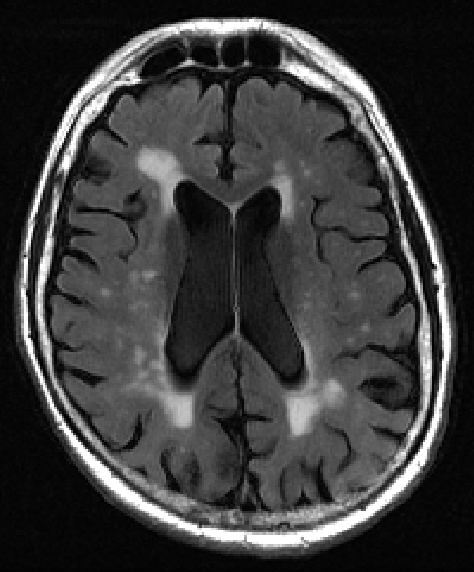
“WHITE MATTER LESION” MAPPING TOOL IDENTIFIES EARLY SIGNS OF DEMENTIA
A new tool for analyzing tissue damage seen on MRI brain scans can detect with more than 70 percent accuracy early signs of cognitive decline, new research shows.