Chemists at the U.S. Department of Energy’s Brookhaven National Laboratory, Stony Brook University (SBU), and their collaborators have uncovered new details of the reversible assembly and disassembly of a platinum catalyst. The new understanding may offer clues to the catalyst’s stability and recyclability.
Tag: Electron Microscopy
Using Ion Beams to Improve Brain Microscopy
Improving the way scientists can see the microscopic structures of the brain can improve our understanding of a host of brain diseases, like Alzheimer’s or multiple sclerosis. Studying these diseases is challenging and has been limited by accuracy of available models.To see the smallest parts of cells, scientists often use a technique called electron microscopy.
Previously Overlooked Algae Toxin Widespread in Southern Indian River Lagoon
Pseudo-nitzschia spp., an algae that produces the neurotoxin domoic acid, can bioaccumulate within food webs causing harm to humans and animals. A molecular study of Florida’s Indian River Lagoon shows this algae was present in 87 percent of the water samples collected. All isolates showed toxicity, and domoic acid was found in 47 percent of surface water samples. As a nursery for many organisms that supports a high amount of biodiversity, the presence of domoic acid could negatively impact the lagoon system.
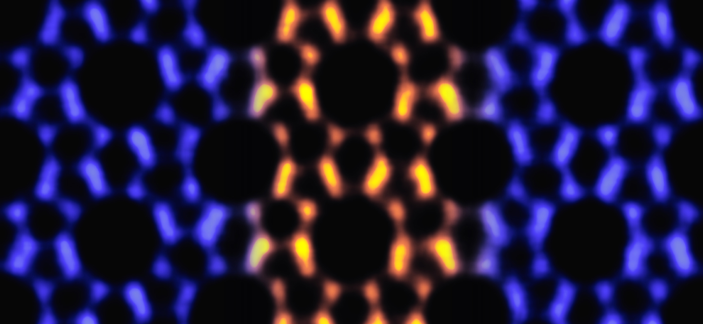
Shining potential of missing atoms
Single photons have applications in quantum computation, information networks, and sensors, and these can be emitted by defects in the atomically thin insulator hexagonal boron nitride (hBN). Missing nitrogen atoms have been suggested to be the atomic structure responsible for this activity, but it is difficult to controllably remove them.
Novel AI-based software enables quick and reliable imaging of proteins in cells
In a cryo-ET experiment, scientists use a transmission electron microscope to obtain 3D images, called tomograms, of the cellular volume containing complex biomolecules.
See-through zebrafish, new imaging method put blood stem cells in high-resolution spotlight
MADISON — For the first time, researchers can get a high-resolution view of single blood stem cells thanks to a little help from microscopy and zebrafish.Researchers at the University of Wisconsin–Madison and the University of California San Diego have developed a method for scientists to track a single blood stem cell in a live organism and then describe the ultrastructure, or architecture, of that same cell using electron microscopy.
Rethinking the rabies vaccine
Rabies virus kills a shocking 59,000 people each year, many of them children. In a new study, researchers from La Jolla Institute for Immunology and Institut Pasteur share a promising path to better vaccine design.
Tracking Pileups on Battery Charging Route to Drive Performance
An understanding of this mechanism could help scientists increase the total amount of energy stored by next-generation lithium-ion batteries.
This crystal impurity is sheer perfection
Scientists at Berkeley Lab and UC Berkeley have developed a nanoparticle composite that grows into 3D crystals. The new 3D-grown material could speed up production and eliminate errors in the mass manufacturing of nanoscale photonics for smart buildings or actuators for robotics.
Mouse brain imaged from the microscopic to the macroscopic level
Researchers at the University of Chicago and the U.S. Department of Energy’s (DOE) Argonne National Laboratory have leveraged existing advanced X-ray microscopy techniques to bridge the gap between MRI (magnetic resonance imaging) and electron microscopy imaging, providing a viable pipeline for multiscale whole brain imaging within the same brain
ORNL’s Sergei Kalinin elected Fellow of the Microscopy Society of America
Sergei Kalinin, a scientist and inventor at the Department of Energy’s Oak Ridge National Laboratory, has been elected a Fellow of the Microscopy Society of America professional society.
ORNL’s superb materials expertise, data and AI tools propel progress
At the Department of Energy’s Oak Ridge National Laboratory, scientists use artificial intelligence, or AI, to accelerate the discovery and development of materials for energy and information technologies.

Science Snapshots From Berkeley Lab – Week of March 29, 2021
India’s Ambitious Clean Energy Goals, a Secret Pathway to Harnessing the Sun for Clean Energy, and a Supersmart Gas Sensor for Asthmatics
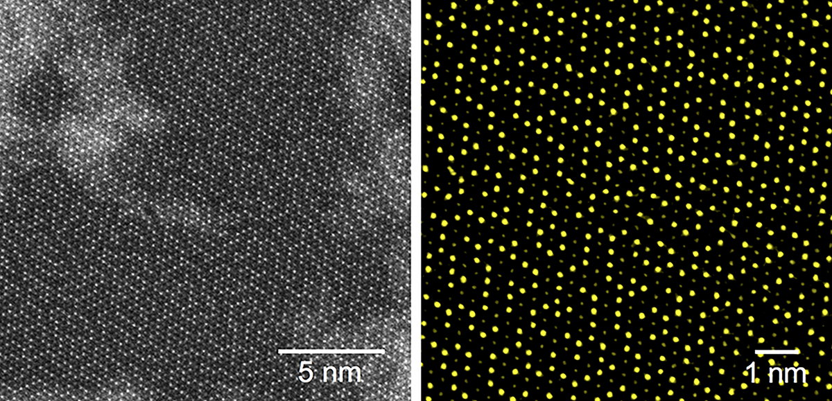
Revealing Nano Big Bang – Scientists Observe the First Milliseconds of Crystal Formation
At Berkeley Lab’s Molecular Foundry, scientists recruited a world-leading microscope to capture atomic-resolution, high-speed images of gold atoms self-organizing, falling apart, and then reorganizing many times before settling into a stable, ordered crystal.
Nikhil Tiwale: Practicing the Art of Nanofabrication
Applying his passions for science and art, Nikhil Tiwale—a postdoc at Brookhaven Lab’s Center for Functional Nanomaterials—is fabricating new microelectronics components.
Study: Turning a coronavirus protein into a nanoparticle could be key to an effective COVID-19 vaccine
One of the proteins on the virus – located on the characteristic COVID spike – has a component called the receptor-binding domain, or RBD, which is its “Achilles heel.” That is, he said, antibodies against this part of the virus have the potential to the neutralize the virus.
October 27, 2020 Web Feature Enabling the Data-Driven Future of Microscopy
An international research team led by PNNL has published a vision for electron microscopy infused with the latest advances in data science and artificial intelligence. Writing a commentary in Nature Materials, the team proposes a highly integrated, autonomous, and data-driven microscopy architecture to address challenges in energy storage, quantum information science, and materials design.
2D Electronics Get an Atomic Tuneup
Scientists at Berkeley Lab have demonstrated a new technique that could improve the performance of atomically thin semiconductors for next-generation electronics such as optoelectronics, thermoelectrics, and sensors.

Pioneering method reveals dynamic structure in HIV
The method reveals that the lattice, which forms the major structural component of the human immunodeficiency virus (HIV), is dynamic. The discovery of a diffusing lattice made from Gag and GagPol proteins, long considered to be completely static, opens up potential new therapies. The method can be applied to biomedical structure.
Peering into Functioning Batteries with Sooyeon Hwang
Using electron microscopes, Hwang—a materials scientist at Brookhaven Lab’s Center for Functional Nanomaterials (CFN)—characterizes the structure and chemistry of operating battery electrode materials.

Story Tips: Molding matter atom by atom and seeing inside uranium particles
Story Tips: Molding matter atom by atom and seeing inside uranium particles, from the Department of Energy’s Oak Ridge National Laboratory
Brookhaven Lab’s Lijun Wu Receives 2020 Chuck Fiori Award
For the past 20 years, Wu has been advancing quantitative electron diffraction to study batteries, catalysts, and other energy materials.
Atomic-scale imaging reveals secret to thin film strength
An international team of scientists and engineers have made a discovery that could further advance the use of ultra-thin zeolite nanosheets, which are used as specialized molecular filters. The discovery could improve efficiency in the production of gasoline, plastics, and biofuels.
Physician-scientist bridges the neurobiology lab to the NICU
As a neonatologist and basic scientist at the University of Chicago Medicine, Tim Sanders, MD, PhD, both provides care for vulnerable infants and studies some of the most fundamental elements of life.