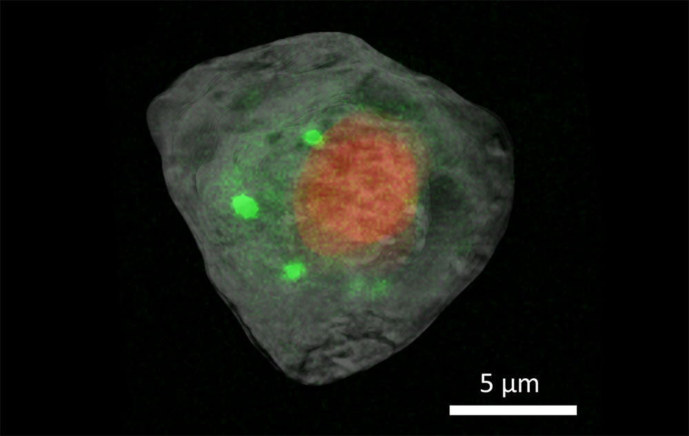
Scientists use a multimodal approach that combines hard X-ray computed tomography and X-ray fluorescence imaging to see the structure and chemical processes inside of a single cell.
news, journals and articles from all over the world.
Scientists use a multimodal approach that combines hard X-ray computed tomography and X-ray fluorescence imaging to see the structure and chemical processes inside of a single cell.

Cell biologist embraces new tools to study human development on the smallest scale
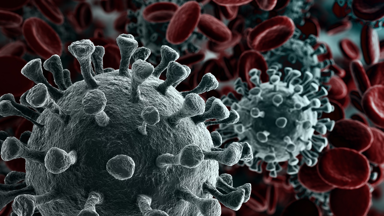
New research from UNC Charlotte’s Center for Computational Intelligence to Predict Health and Environmental Risks has found that the two most prevalent strains of the virus that cause COVID-19, SARS-CoV-2 variants BA.2.86 and JN.1, are not significantly better than their predecessor Omicron at evading immune responses and causing infections despite having a high number of mutations compared to previous variants.
New drug inspired by images that captured how bacteria block antibiotic activity
In October 2023, the Advanced Photon Source (APS), a U.S. Department of Energy (DOE) Office of Science user facility at DOE’s Argonne National Laboratory, officially launched a new initiative to expand biological and environmental research at the world leading X-ray and analysis facility.
Antibody therapies are only effective if the antibodies do what we want them to do. This research can help scientists determine if an antibody is likely to stick to something other than the intended target, which should lessen the amount of time wasted with overly sticky antibodies.
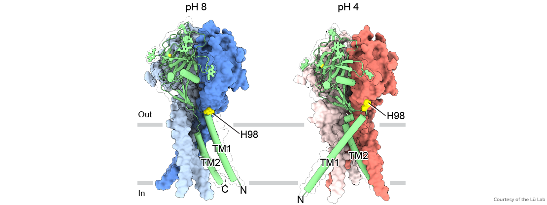
Scientists at Brookhaven Lab have determined the atomic-level structure of a zinc-transporter protein, a molecular machine that regulates levels of this crucial trace metal micronutrient inside cells. The structure reveals how the cellular membrane protein shifts its shape to move zinc from the environment into a cell, and temporarily blocks this action automatically when zinc levels inside the cell get too high.
Researchers from Argonne, Michigan State University and Northwestern University used Argonne’s Bionanoprobe beamline to look at the concentration of zinc in egg cells.
In a cryo-ET experiment, scientists use a transmission electron microscope to obtain 3D images, called tomograms, of the cellular volume containing complex biomolecules.
Scientists at St. Jude Children’s Research Hospital have found that human ribosomes decode mRNA slower than bacteria, with implications for drug development.
Scientists at Brookhaven National Laboratory have produced the first atomic-level structure of an enzyme that selectively cuts carbon-hydrogen bonds—the first and most challenging step in turning simple hydrocarbons into more useful chemicals. The detailed atomic level “blueprint” suggests ways to engineer the enzyme to produce desired products.
The cystic fibrosis transmembrane conductance regulator has been studied for years but the combined efforts of St. Jude Children’s Research Hospital and Rockefeller University have yielded new insights.
A group of researchers have used the Advanced Photon Source to look at monoclonal antibodies to subvert the “shield” of the Lassa virus, potentially paving the way for new therapies.
Scientists at St. Jude Children’s Research Hospital captured the 3D structure of SPOP, revealing how mutations in previously unappreciated regions fuel cancer.
A new study led by scientists at La Jolla Institute for Immunology (LJI) shows how ideal antibodies against SARS-CoV-2 hit their marks. Now scientists are looking at how we might harness their power in new antibody therapeutics and even more effective COVID-19 vaccines.
Researchers from Paul Scherrer Institute PSI and ETH Zurich have discovered how proteins in the cell can form tiny liquid droplets that act as a smart molecular glue.
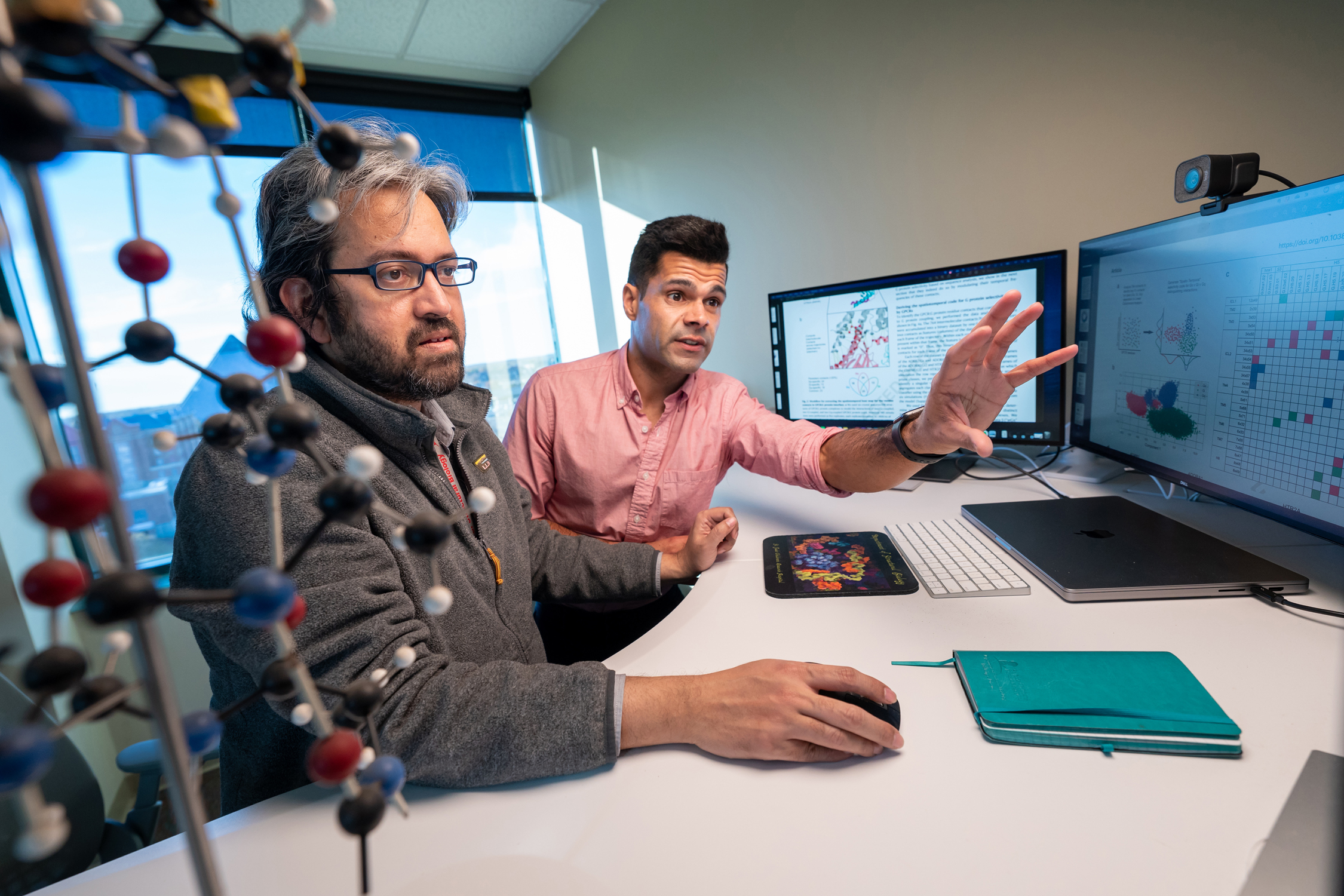
By simulating molecular dynamics, scientists from St. Jude Children’s Research Hospital revealed how the selectivity or promiscuity of GPCR coupling relies on the location and duration of intermolecular interactions.
The LJI team plans to use their new map of the Lassa virus surface glycoprotein to design a much-needed vaccine.
The World Health Organization says monkeypox is a global health emergency. Scientists use ultrabright X-ray beams and diffraction imagery to understand how poxviruses behave. This can accelerate development of critical vaccines and treatments for monkeypox and other poxviruses.
A new study on rubisco, a photosynthetic enzyme thought to be the most abundant protein on the planet, shows that proteins can change their structural arrangement with surprising ease. The findings reveal the possibility that many of the proteins we thought we knew actually exist in other, unknown shapes.
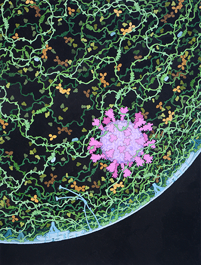
During the annual ACA meeting, David Goodsell, of Scripps Research and RCSB Protein Data Bank, will discuss his use of artistic methods to visualize biological data. His presentation, “Art as a Tool for Structural Biology,” takes place Tuesday. Traditional artistic mediums, like painting, provide the freedom necessary to illustrate cells, and Goodsell is using illustrations to lay the foundation for computational modeling of whole cells. He also creates brightly colored, cartoonlike graphics with nonphotorealistic computer graphics methods to highlight the overall shape of molecules and how they assemble and interact.
A research team led by Caltech spent nearly 20 years determining X-ray structures, one by one, to create a map of the nuclear pore complex, one of the largest and most complex pieces of cellular machinery.
Rabies virus kills a shocking 59,000 people each year, many of them children. In a new study, researchers from La Jolla Institute for Immunology and Institut Pasteur share a promising path to better vaccine design.
GRAND RAPIDS, Mich. (March 21, 2022) — When something goes wrong during DNA replication, cells call their own version of 911 to pause the process and fix the problem — a failsafe that is critical to maintaining health and staving off disease.
Argonne researchers are mapping the complex tangle of the brain’s connections — a connectome — by developing applications that will find their stride in the advent of exascale computing.
Dana Pe’er, PhD, computational biologist and lab head at Memorial Sloan Kettering Cancer Center’s (MSK) Sloan Kettering Institute (SKI), is one of 33 biomedical researchers named a Howard Hughes Medical Institute (HHMI) investigator today.
An experiment to study gravity at the quantum scale, insights into an antibiotic-building enzyme, and the backstory of an incredible new protein prediction algorithm are featured in this month’s roundup of science highlights.
Argonne, industry and academia collaborate to bring innovative AI and simulation tools to the COVID-19 battlefront.
Naked RNA molecules are too floppy for high-res 3D imaging, but a system developed at SLAC and Stanford fixes that. It reveals detailed RNA structures from a pond scum critter and COVID-19 virus.
Naked RNA molecules are too floppy for high-res 3D imaging, but a system developed at SLAC and Stanford fixes that. It reveals detailed RNA structures from a pond scum critter and COVID-19 virus.
Scientists using the Advanced Photon Source have discovered that a drug used to fight tumors in animals might be effective against many types of coronaviruses, including SARS-CoV-2.
Scientists from three national labs have published a comprehensive study that – alongside other recent, complementary studies of coronavirus proteins and genetics – represents the first step toward developing treatments for COVID-19.
Two teams of researchers using the Advanced Photon Source identified existing drugs — one used to treat cancer, the other an anti-seizure medication — that may work as treatments for COVID-19.
Using DNA, scientists organized bioactive proteins in desired 2-D and 3-D ordered arrays—promising for structural biology, biomedicine, and more.
This study investigates how the nucleocapsid protein, or N protein, of the SARS-CoV-2 virus packages the viral genome.
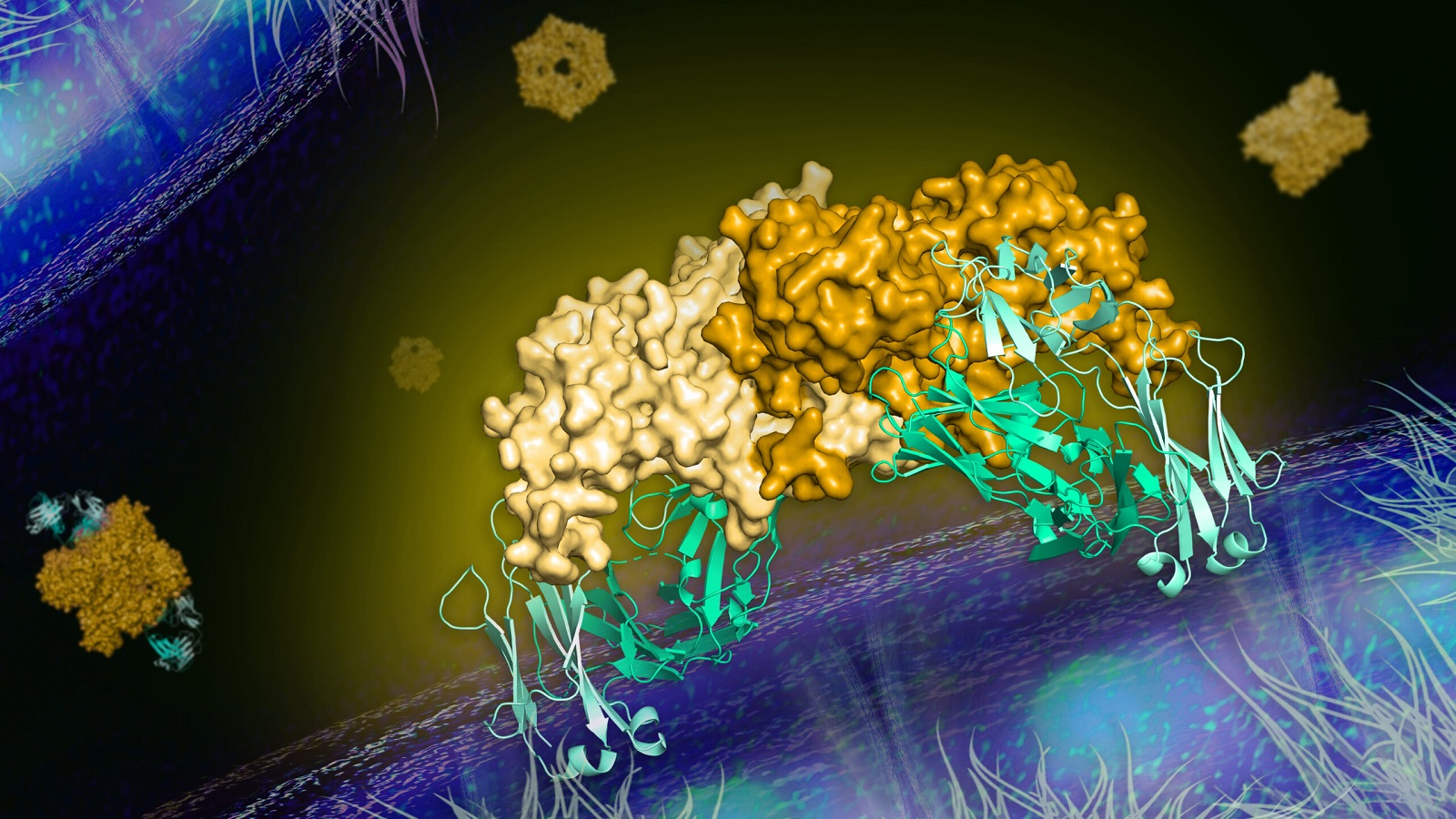
Researchers have developed antibodies that can bind to phosphohistidine, an unstable molecule that’s linked to cancer. To learn how the two bind together, the team turned to the powerful X-rays at Argonne’s Advanced Photon Source. These new insights into its structure will help scientists design better antibodies for potential treatments.
Scientists from around the world will come together virtually to celebrate the 50th anniversary of a key piece of the infrastructure for sharing scientific knowledge: the Protein Data Bank (PDB). The event will be hosted by the American Society for Biochemistry and Molecular Biology on May 4–5, 2021.

As the fight against COVID-19 continues, scientists have turned to an unlikely source for a potentially effective treatment: tiny antibodies naturally generated by llamas.
New Brunswick, N.J. (March 3, 2021) – The 3D structures of more than 1,000 SARS-CoV-2 coronavirus proteins are freely available from the RCSB Protein Data Bank headquartered at Rutgers University–New Brunswick. The data bank reached the milestone this week, with 1,018 proteins as…
In developing therapies for hard-to-treat breast and ovarian cancers in patients with BRCA gene mutations, scientists aim to identify ways to keep cancer cells from using DNA break repair pathways. New findings demonstrate a previously-unknown capability for polymerase theta (pol theta) – a key enzyme in this repair function – that shows promise as a new avenue for treatment development.
The research team used the Advanced Photon Source to confirm an effective antibody that prevents the dengue virus from infecting cells in mice, and may lead to treatments for this and similar diseases.

A team of biologists who banded together to support COVID-19 science determined the atomic structure of a coronavirus protein thought to help the pathogen evade and dampen response from human immune cells. The structural map has laid the groundwork for new antiviral treatments and enabled further investigations into how the newly emerged virus ravages the human body.
Argonne scientists and research facilities have made a difference in the fight against COVID-19 in the year since the first gene sequence for the virus was published.
More than a decade of virus research at the APS laid the groundwork for more effective COVID-19 vaccines and helped speed their rapid development.
The APS has been a powerful tool in the battle against the novel coronavirus, contributing more information about the structure of the virus to the International Protein Databank than any other light source in the United States.
New Brunswick, N.J. (Dec. 3, 2020) – Stephen K. Burley, director of the RCSB Protein Data Bank headquartered at Rutgers University–New Brunswick, is available for interviews on how the bank’s 50 years of data on the 3D biomolecular structures of life and artificial intelligence can lead…
Research teams from across the United States are using a multitude of techniques to study the SARS-CoV-2 virus using the Advanced Photon Source from their homes and institutions.
Argonne scientists, working as part of a national consortium of structural genomics experts, have greatly increased our knowledge of the virus that causes COVID-19.

Argonne computational resources supported the largest comprehensive analysis of COVID-19 genome sequences in the U.S. and helped corroborate growing evidence of a protein mutation.
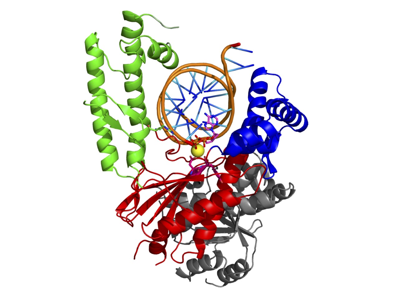
GRAND RAPIDS, Mich. (Nov. 4, 2020) — For the first time, scientists have visualized a new class of molecular gates that maintain pH balance within brain cells, a critical function that keeps cells alive and helps prevent stroke and other brain injuries.