There is a clear relationship between alcohol and neurodegeneration; for example, an alcohol use disorder correlates with a higher risk of Alzheimer’s disease. It is unclear, however, whether individual differences in brain structure and connectivity are risk factors for, or consequences of, alcohol use. New research exploring this relationship will be shared on Wednesday, 26 June 2024 at the 47th annual scientific meeting of the Research Society on Alcohol (RSA) in Minneapolis, Minnesota.
Tag: Brain Cells
Super-Chilled Brain Cell Molecules Reveal How Epilepsy Drug Works
By super cooling a molecule on the surface of brain cells down to about minus 180 degrees Celsius — nearly twice as cold as the coldest places in Antarctica — scientists at Johns Hopkins Medicine say they have determined how a widely-used epilepsy drug works to dampen the excitability of brain cells and help to control, although not cure, seizures.
‘MUSIC map’ reveals some brain cells age faster and are more prevalent in Alzheimer’s
Engineers at the University of California San Diego have discovered that some brain cells age more rapidly than others, and they are disproportionately abundant in individuals afflicted with Alzheimer’s disease. Additionally, researchers observed sex-specific differences in the aging process of certain brain cells, with the female cortex exhibiting a higher ratio of “old” oligodendrocytes to “old” neurons compared to the male cortex.

Cedars-Sinai Study Details Workings of Short-Term Memory
Cedars-Sinai investigators have discovered how brain cells responsible for working memory—the type required to remember a phone number long enough to dial it—coordinate intentional focus and short-term storage of information.
Saturated fat may interfere with creating memories in aged brain
New research hints at a few ways fatty foods affect cells in the brain, a finding that could help explain the link between a high-fat diet and impaired memory – especially as we age.
UC Irvine to lead multi-institutional study of single-cell vulnerabilities to Alzheimer’s disease
The University of California, Irvine has received a five-year, $10 million grant from the National Institutes of Health to lead a multi-institutional study of specific brain cell vulnerabilities to abnormal tau protein deposits in regions affected in the early stages of Alzheimer’s disease.
Mount Sinai Researchers Identify Potential New Treatment for Those Who Act Out Their Dreams While Sleeping
Experts say medication commonly used to treat insomnia may also be a therapeutic option for the condition known as REM sleep behavior disorder
UC San Diego, Salk and Others Seek to Map the Human Brain Over a Lifetime
With a $126 million grant from the National Institutes of Health, a multi-institution team of researchers at UC San Diego, Salk Institute and elsewhere has launched a new Center for Multiomic Human Brain Cell Atlas to describe human brain cells in unprecedented detail over a lifetime.
Scientists Discover Cellular Stress Enzyme That Might Play Key Role in Neurodegenerative Diseases Such as ALS
An enzyme called MARK2 has been identified as a key stress-response switch in cells in a study by researchers at Johns Hopkins Bloomberg School of Public Health.
Research News Tip Sheet: Story Ideas From Johns Hopkins Medicine
During the COVID-19 pandemic, Johns Hopkins Medicine Media Relations is focused on disseminating current, accurate and useful information to the public via the media. As part of that effort, we are distributing our “COVID-19 Tip Sheet: Story Ideas from Johns Hopkins” every other Tuesday.
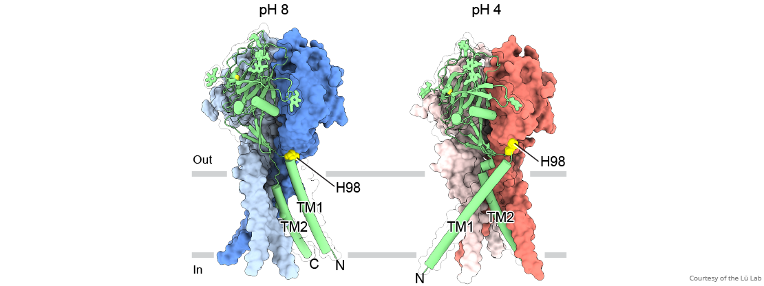
Near-atomic ‘maps’ reveal structure for maintaining pH balance in cells
GRAND RAPIDS, Mich. (Nov. 4, 2020) — For the first time, scientists have visualized a new class of molecular gates that maintain pH balance within brain cells, a critical function that keeps cells alive and helps prevent stroke and other brain injuries.

Research News Tip Sheet: Story Ideas From Johns Hopkins Medicine
During the COVID-19 pandemic, Johns Hopkins Medicine Media Relations is focused on disseminating current, accurate and useful information to the public via the media. As part of that effort, we are distributing our “COVID-19 Tip Sheet: Story Ideas from Johns Hopkins” every other Tuesday.
Microglia Might Lessen Seizure Severity in Epilepsy
Article title: Microglia depletion exacerbates acute seizures and hippocampal neuronal degeneration in mouse models of epilepsy Authors: Mei Liu, Lijuan Jiang, Min Wen, Yue Ke, Xiangzhen Tong, Weiyuan Huang, Rongqing Chen From the authors: “Our study indicates that microglia are innately ready…
Brain’s Immune Cells Promising Cellular Target for Therapeutics
Inspired by the need for new and better therapies for neurodegenerative diseases, Rutgers University researchers are exploring the link between uncontrolled inflammation within the brain and the brain’s immune cells, known as microglia, which are emerging as a promising cellular target because of the prominent role they play in brain inflammation. In APL Bioengineering, the group highlights the design considerations and benefits of creating therapeutic nanoparticles for carrying pharmacological factors directly to the sites of the microglia.
Microglia Might Lessen Seizure Severity in Epilepsy
New research in mice highlights the potential protective effect of microglia—a type of non-neuronal cell in the brain—against overactivation of the central nervous system during acute epileptic seizures. The study is published in the American Journal of Physiology-Cell Physiology.
Cumulative Effects of Long Term Alcohol on Brain Function
Functional MRI (fMRI), a type of scan that measures brain activity, has enabled study of the impact of alcohol on brain function. This type of imaging allows brain activity to be assessed while participants are at rest, performing a simple task like tapping a finger, or doing a complex cognitive task like a memory task or decision-making. It works by detecting the change in blood flow that occurs when brain cells (or neurons) in different parts of the brain are activated. Blood flow provides the energy and oxygen needed for brain cells to activate, and it is this exchange of oxygen that is measured using fMRI and is reflected by brain blood flow. Complicated physics are involved in determining the profile of blood flow when a part of the brain is activated, and studies have shown that the time course of these changes – known as the hemodynamic response function (HRF) – is affected by acute alcohol consumption. However, the effects of heavy chronic (long-term) alcohol consumption on HRF
Think Before You Drink: The Brain Plays a Role in Alcoholic Fatty Liver Disease
New research shows that two brain proteins help regulate fat accumulation in the liver associated with excessive alcohol consumption, specifically binge drinking. The study is published ahead of print in the American Journal of Physiology—Endocrinology and Metabolism.