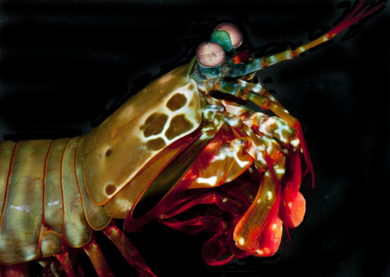
Irvine, Calif., Aug. 17, 2020 – University of California, Irvine materials scientists are learning about resilience from the mantis shrimp. The ancient crustaceans are armed with two hammerlike raptorial appendages called dactyl clubs that they use to bludgeon and smash their prey. These fists, able to accelerate from the body at over 50 mph, deliver powerful blows yet appear undamaged afterward.
Tag: Microscopy
UIC physicist earns innovation award from Microscopy Today
Groundbreaking approach in high-resolution electron energy loss spectroscopy has been judged to be one of the 10 best microscopy innovations in the 2020 Microscopy Today Innovation Award competition.
New Research Reveals Antifungal Symbiotic Peptide In Legume
Danforth Center scientists, Dilip Shah, PhD, Siva Velivelli, PhD, Kirk Czymmek, PhD, and their collaborators at the Pacific Northwest National Laboratory have identified a sub class of peptides in the nodules of the legume, Medicago truncatula that proved effective in inhibiting growth of the fungus causing gray mold.
Pioneering method reveals dynamic structure in HIV
The method reveals that the lattice, which forms the major structural component of the human immunodeficiency virus (HIV), is dynamic. The discovery of a diffusing lattice made from Gag and GagPol proteins, long considered to be completely static, opens up potential new therapies. The method can be applied to biomedical structure.
Sorting and secreting insulin by expiration date
The age of insulin parcels may matter, researchers say, when it comes to diagnosing and treating diabetes.
Story Tips: Predicting fire risk, solid state stability check and images in a flash
ORNL Story Tips: Predicting fire risk, solid state stability check and images in a flash
Faster processing makes cutting-edge fluorescence microscopy more accessible
Scientists at NIBIB have developed new image processing techniques for microscopes that can reduce post-processing time up to several thousand-fold.
Good vibrations show how water works
Researchers demonstrated new ways to use electron microscopy to study liquids at high resolution. They used this technique to examine how nuclei in liquids and molecules vibrate at multiple length scales. This work can lead to new ways for scientists to describe liquids, the interfaces between fluids, and materials labeled with isotopes.
Eye Injury Sets Immune Cells On Surveillance To Protect The Lens
The discovery further challenges the accepted scientific dogma that the lens is shut out from the immune protection.
After Turning Microorganisms Into Art, Student Helps NASA Study Origins of Life Through Algae (Video)
Rutgers student Julia Van Etten, whose @Couch_Microscopy Instagram page garnered more than 25,000 followers by showcasing microorganisms as art, is now working with NASA on research into how red algae can help explain the origins of life on Earth.
Seeing starch: Novel technique enables gentle observation of biofuel materials
Scientists at the Department of Energy’s Oak Ridge National Laboratory have developed a new method to peer deep into the nanostructure of biomaterials without damaging the sample. This novel technique can confirm structural features in starch, a carbohydrate important in biofuel production.
Like a Microscopic Mars Rover, a New Technique Tracks Individual Protein Movement on Live Cells
The piece of gold that Richard Taylor was thrilled to track down weighed less than a single bacterium. Taylor, a postdoctoral fellow at the Max Planck Institute, was working to follow individual nanogold-labeled molecules that move just nanometers, billionths of a meter.
Channeling Electrons for Ultrafast Spin Conductivity
Scientists used unique scanning probe microscopy and spectroscopy techniques to control how electrons moved on the surface of a bismuth-based material (Bi2Te2Se).
New Algorithms Shed Light on Molecules’ Structure and Motion in Cells
To understand why very large molecules behave the way they do inside cells, scientists must first understand the relationship between these molecules’ structure and motion. Engineers created algorithms that provide the physics backbone for a new “microscope in a computer.”
Science Snapshots – microbiome matchmakers, solid-liquid interfaces, undersea earthquakes
Science Snapshots from Berkeley Lab
Advanced microscopy reveals unusual DNA structure
An advanced imaging technique reveals new structural details of S-DNA, ladder-like DNA that forms when the molecule experiences extreme tension. This work conducted at Sandia National Laboratories and Vrije University in the Netherlands provides the first experimental evidence that S-DNA contains highly tilted base pairs.
UCI scientists reveal mechanism of electron charge exchange in molecules
Irvine, Calif., Oct. 14, 2019 – Researchers at the University of California, Irvine have developed a new scanning transmission electron microscopy method that enables visualization of the electric charge density of materials at sub-angstrom resolution. With this technique, the UCI scientists were able to observe electron distribution between atoms and molecules and uncover clues to the origins of ferroelectricity, the capacity of certain crystals to possess spontaneous electric polarization that can be switched by the application of an electric field.