The Science
The Impact
Detecting cancer at an early stage is one of the most critical factors to successfully treating it and preventing death. Developing a low-cost, high-throughput microscopy technique that can sense changes in large molecules in living cells is a key step towards this goal. Using PWS’s high resolution with the new algorithm opens the door for more accurate, more efficient early-stage cancer screenings. Now it may be possible to detect seven different human cancers with minimal false positives: lung, colon, ovarian, esophageal, pancreatic, thyroid, and prostate.
Summary
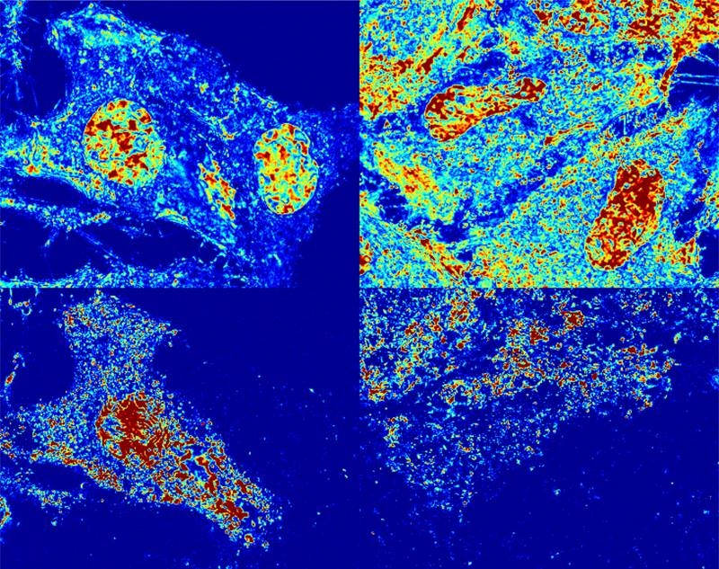
The finite time-difference time-domain code, Angora, is a computational microscopy code that models the four components of an optical imaging system (illumination, scattering, collection, and refocusing). It then synthesizes microscope images to validate theoretical results and predict scattering behaviors. Simulations with Angora were critical in this project to validate the analytic theory developed to extract physical properties from the measured spectral interference. The team then applied PWS to explore the nanoscale structural and dynamic changes of stem cells. Experiments probed the spatio-temporal response of macromolecular assemblies to ultraviolet (UV) light. Scientists discovered a near instantaneous burst of motion that occurs early in process of UV-induced cell death. This process called cellular paroxysm was found to occur synchronously within a cell, as far as 30 micrometers apart in under 35 milliseconds, but asynchronously between cells. The team notes that this effect may play a role in the earliest stages of a unique form of cell death. However, the exact mechanism of cell death is unclear. The team also developed a machine learning model to classify cells based solely on physical properties of the nucleus with an accuracy of 80% and indicated that potential improvements could come by way of larger datasets.
Funding
The research was supported by the National Institutes of Health, National Cancer Institute, National Science Foundation, and the NIH Chemistry of Life Processes Predoctoral training program. Time on the Argonne Leadership Computing Facility (ALCF), a DOE scientific user facility, through the INCITE program.
Original post https://alertarticles.info

