Watching and measuring what happens in tissues inside the human embryo is currently not possible, and it is still very difficult to do this in mammalian models like mice. Because humans and the fruit fly Drosophila share so many biological similarities, researchers from Columbia Engineering and Syracuse University decided to tackle this problem by focusing on fruit flies. In a paper published online May 29 in PNAS, the team reports that they can predict when the tissue will begin to rapidly flow just by looking at cell shapes in the tissue.
“Thanks to earlier theoretical work from our colleagues at Syracuse, we thought we might be able to learn something about whether the embryonic tissues are solid or fluid by just looking at the shapes of cells in the tissue,” says the study’s lead PI Karen Kasza, Clare Boothe Luce Assistant Professor of Mechanical Engineering. “So we decided to try this in the fly. We’re really excited about our results, which could reveal fundamental mechanisms underlying human development and point to where things can go wrong, causing birth defects.”
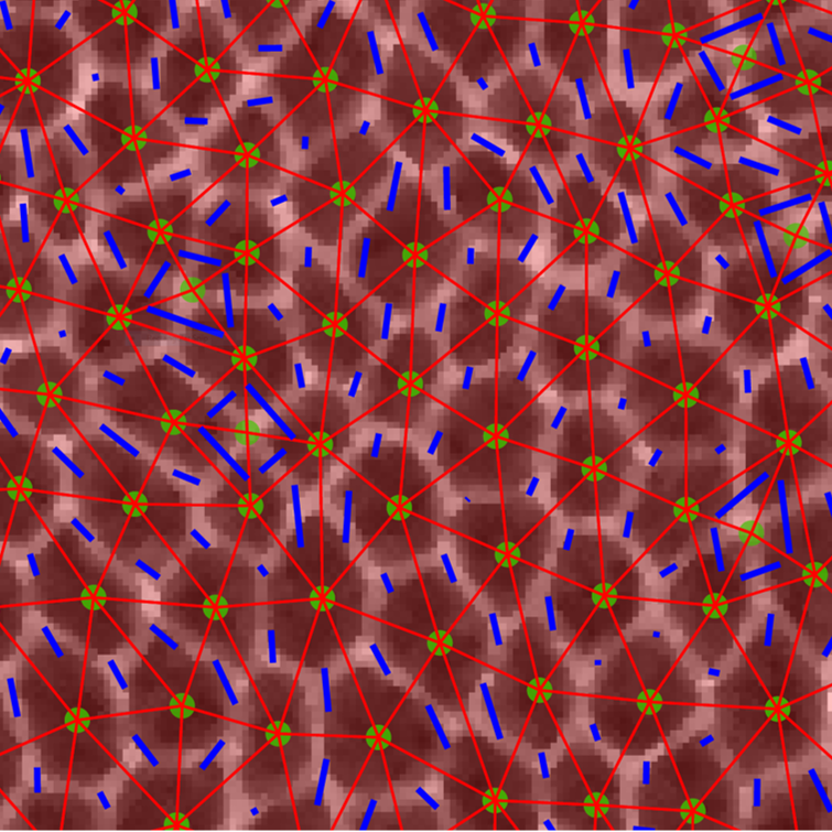
The challenge was how to apply traditional engineering approaches to measure the mechanical properties of cells and tissues inside the flies’ tiny embryos to see which tissues behave like solids, maintaining their shape and resisting flow, and which tissues behave like fluids, flowing easily and changing shape. The researchers used high-resolution confocal fluorescence imaging to take movies of embryonic development in which they could see in great detail the shape and packings of cells in tissues inside the fly embryo. They focused on a very fast-moving developmental event in which the embryonic tissue rapidly changes shape to elongate the head-to-tail body axis of the fly (something that also happens in most animal embryos).
By combining experimental studies in the fruit fly embryo at Columbia with theoretical modeling approaches at Syracuse, the researchers demonstrated that the shapes and alignment of cells within tissues can help to both explain and predict how tissues change shape during development and how defects in these processes can result in abnormalities in embryo shape. “It was a fantastic collaboration between experiment and theory,” Kasza observes.
A surprise for the researchers was that they could anticipate when the tissue would begin to flow by looking at cell shapes in the tissue without any adjustable parameters in the theoretical model. But, unlike previous studies and predictions, they needed to include a new parameter—anisotropy—that described the alignment of cells within the tissue because the forces acting on and in the tissue were highly anisotropic, or varied along different directions in the tissue. What they found particularly interesting was that their findings suggest that embryonic tissue seems to become more fluid-like just before the onset of the rapid tissue flows during body axis elongation.
“This is really exciting” says Kasza, “because it suggests that the mechanical properties of the cells might be regulated biologically during embryonic development, i.e. in the genetic instructions encoded in DNA, to make it easier for tissues to change shape dramatically during brief time windows during development. This adds to a growing body of research revealing that mechanics is really crucial to understanding life.”
The team is now looking at how the instructions for changes in tissue fluidity are genetically encoded. They are also exploring the mechanical properties of tissues to build quantitative models of tissue morphogenesis that will enable them to predict, design, build, and control tissue shape and tissue movements, both in developing embryos and in engineered tissues in the lab.
About the Study
The study is titled: “Anisotropy links cell shapes to tissue flow during convergent extension.”
Authors are: Xun Wang 1; Matthias Merkel 2,3; Leo B. Sutter 2; Gonca Erdemci-Tandogan 2; M. Lisa Manning 2; Karen E. Kasza 1.
1 Department of Mechanical Engineering, Columbia Engineering
2 Department of Physics and BioInspired Institute, Syracuse University
3 Aix Marseille Univ, Université de Toulon, CNRS, CPT, Turing Center for Living Systems, Marseille, France
Xun Wang, Matthias Merkel, and Leo B. Sutter contributed equally on this work.
The study was supported by the National Science Foundation CMMI 1751841 to K.E.K., DMR-1352184 and POLS-1607416 to M.L.M, and DMR-1460784 (REU) to L.B.S. M.L.M., M.M., and G.E.T. acknowledge support from Simons Grant No. 446222 and 454947, and NIH R01GM117598. K.E.K. holds a Burroughs Wellcome Fund Career Award at the Scientific Interface, Clare Boothe Luce Professorship, and Packard Fellowship.
The authors declare no financial or other conflicts of interest.
###
LINKS:
Paper: https://www-pnas-org.ezproxy.cul.columbia.edu/content/early/2020/05/27/1916418117/tab-figures-data
DOI: 10.1073/pnas.1916418117
https://www.pnas.org/
http://engineering.columbia.edu/
https://engineering.columbia.edu/faculty/karen-kasza
https://thecollege.syr.edu/people/faculty/manning-m-lisa/
https://me.columbia.edu/
###
Columbia Engineering
Columbia Engineering, based in New York City, is one of the top engineering schools in the U.S. and one of the oldest in the nation. Also known as The Fu Foundation School of Engineering and Applied Science, the School expands knowledge and advances technology through the pioneering research of its more than 220 faculty, while educating undergraduate and graduate students in a collaborative environment to become leaders informed by a firm foundation in engineering. The School’s faculty are at the center of the University’s cross-disciplinary research, contributing to the Data Science Institute, Earth Institute, Zuckerman Mind Brain Behavior Institute, Precision Medicine Initiative, and the Columbia Nano Initiative. Guided by its strategic vision, “Columbia Engineering for Humanity,” the School aims to translate ideas into innovations that foster a sustainable, healthy, secure, connected, and creative humanity.
Original post https://alertarticles.info

