To overcome these limitations, a collaboration between Institut Fresnel at Aix Marseille University and the Centre de Biologie Intégrative at Toulouse University in France led to the development of a new type of microscope called Extended Depth-of-Field Random Illumination Microscopy (EDF-RIM). This innovative approach combines the benefits of super-resolution microscopy with an extended depth-of-field detection scheme, allowing for the capture of entire volumes in a single image.
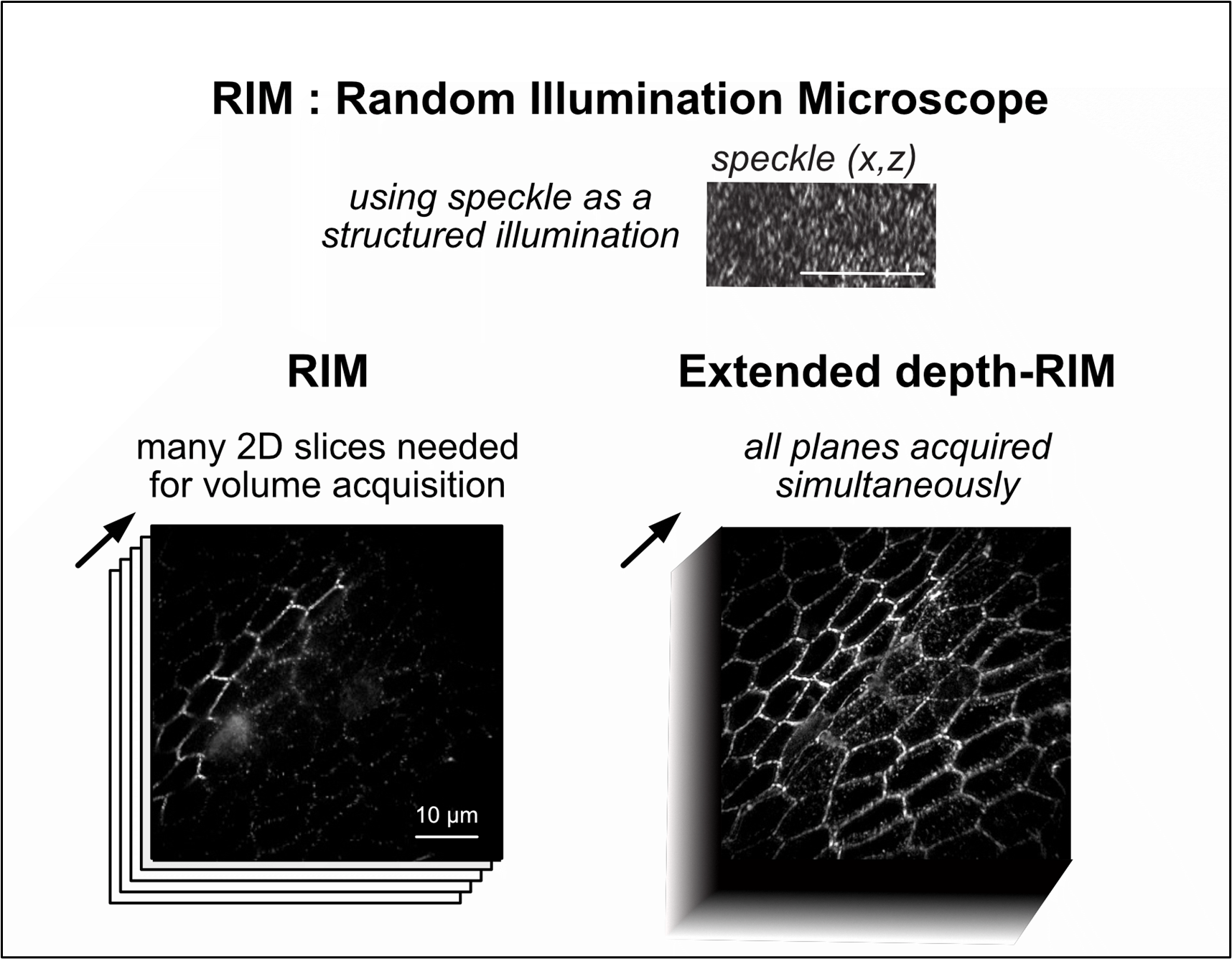
EDF-RIM is built upon the foundation of Random Illumination Microscopy (RIM), a super-resolution technique that utilizes random speckle patterns for illumination. These patterns are statistically insensitive to scattering or aberrations. RIM uses multiple images taken under different speckle patterns to generate a super-resolved image through a numerical algorithm that analyzes the variance of the images. This unique approach makes RIM particularly well-suited for 3D imaging.
EDF-RIM addresses the limitation of needing multiple images per slice by incorporating an extended depth-of-field (EDF) detection system. This system rapidly scans the focal plane, effectively projecting the entire 3D volume onto a single image. This dramatically reduces the time needed for image acquisition and significantly minimizes light exposure.
The EDF-RIM system has been shown to deliver significant advantages over conventional EDF microscopy, including improved contrast and a two-fold enhancement in resolution. However, the projection nature of EDF-RIM means that axial information is lost. For samples where fluorescence is distributed over a curved surface, such as drosophila epithelium, the researchers have developed a method to estimate the surface topography. This allows for the reconstruction of a laterally super-resolved 3D image with accurate representation of the object’s shape.
EDF-RIM represents a significant advancement in fluorescence microscopy, offering a more efficient and powerful way to image large and complex 3D structures. This technology holds tremendous promise for advancing our understanding of biological processes, especially in areas where speed, resolution, and minimal light exposure are critical factors.
###
References
DOI
Original Source URL
https://doi.org/10.1038/s41377-024-01612-0
Funding information
Agence Nationale de la Recherche (ANR-18-CE13-028, ANR-20-CE45-0024, ANR-22-CE13-0039, ANR-22-CE42-0010, ANR-22-CE42-0026); Institut Carnot star (3D-RIM).
This project is funded by the « France 2030 » investment plan managed by the French National Research Agency (ANR-16-CONV-0001, ANR-21-ESRE-0002), and from Excellence Initiative of Aix-Marseille University – A*MIDEX.
About Light: Science & Applications
The Light: Science & Applications will primarily publish new research results in cutting-edge and emerging topics in optics and photonics, as well as covering traditional topics in optical engineering. The journal will publish original articles and reviews that are of high quality, high interest and far-reaching consequence.

