These tools can shed light on the biological processes occurring at the macromolecular and subcellular levels, which usually remain hidden from sight as most tissue is opaque to visible radiation. In APL Photonics, by AIP Publishing, researchers from the Leibniz Institute of Photonic Technology in Germany created a particularly narrow endoscope made of single hair-thin optical fibers that uses holographic methods to reconstruct images of macroscopic objects placed in front of the far end of the endoscope.
“We were positively surprised that the imaging quality was well-maintained at larger imaging distances, even for objects placed at a half meter from the endoscope,” said author Ivo Leite. “We expected that the low number of photons collected in this range would give rise to much higher detection noise.”
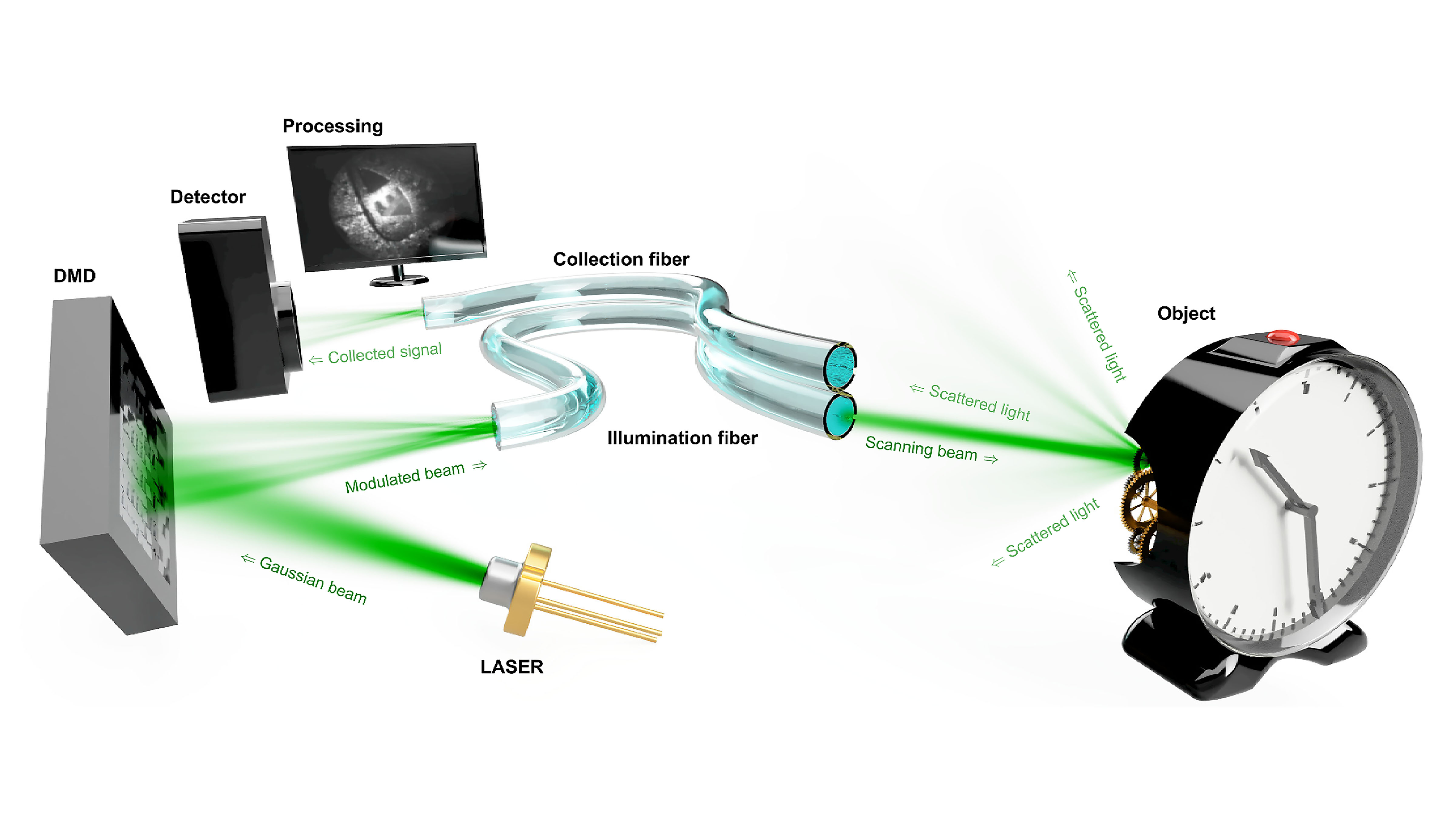
Efforts in imaging through multimode-fiber endoscopes previously focused on working distances typically smaller than 20 micrometers to resolve micrometer-scale details. This limits the field of view to the size of the fiber core.
The researchers brought the imaging operation to the observation of macroscopic objects, which can be placed far away from the endoscope. Researchers increased the imaging performance in terms of image definition to 100,000 pixels per image frame, an order of magnitude larger than previous holographic endoscopes and reaching the definition of modern video endoscopes.
Their efforts pave the way for bringing this class of minimally invasive endoscopes to clinical applications. The macroscopic imaging modality shown in this study will be essential to analyze biological samples at the tissue scale — just as conventional clinical endoscopes do — as well as to guide the instrument insertion.
Once a region of interest is identified, the hologram sequence displayed by the spatial light modulator can be updated to switch the imaging modality and perform observations at the cellular and subcellular levels.
“The potential for such flexibility in imaging operation through the same unmodified endoscope is a unique feature that, we believe, holographic endoscopes could soon offer,” said author Tomas Cizmar.
The researchers’ light control methods could be used to deliver practically any type of photonics tool through a hair-thin endoscope, which could have applications in a range of areas, such as optical transfection, subcellular laser surgery, and laser-assisted microfabrication.
###
The article “Observing distant objects with a multimode fiber-based holographic endoscope” is authored by Ivo T. Leite, Sergey Turtaev, Dirk E. Boonzajer Flaes, and Tomas Cizmar. The article will appear in APL Photonics on March 30, 2021 (DOI: 10.1063/5.0038367). After that date, it can be accessed at https://aip.scitation.org/doi/10.1063/5.0038367.
ABOUT THE JOURNAL
APL Photonics is the dedicated home for open access multidisciplinary research from and for the photonics community. The journal publishes fundamental and applied results that significantly advance the knowledge in photonics across physics, chemistry, biology and materials science. See https://aip.scitation.org/journal/app.
###

