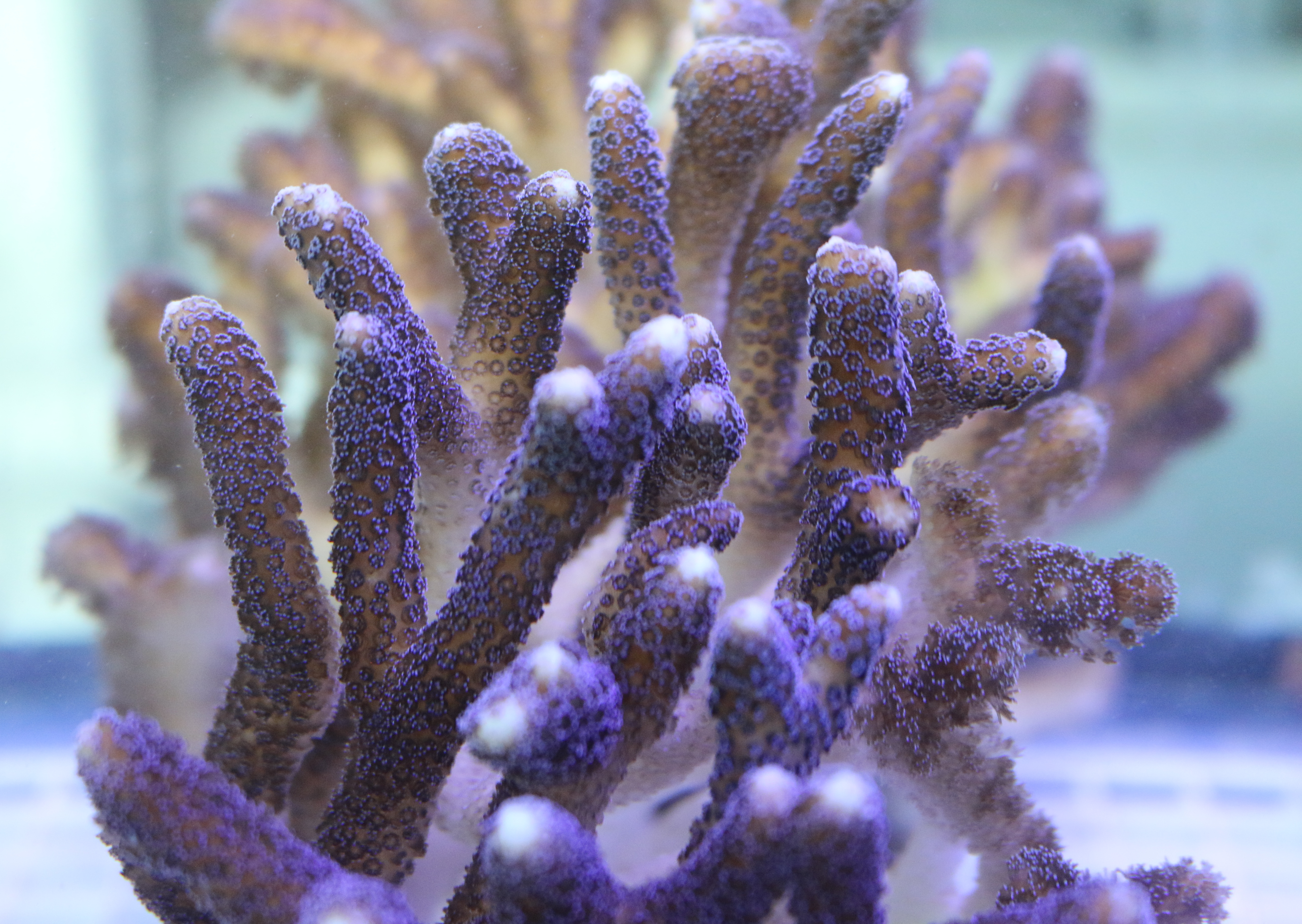
A low-cost and rapid 3D technique is helping scientists to gain insight into the colony- and community-level dynamics of the poorly understood stony coral tissue loss disease responsible for widespread coral death throughout the Tropical Western Atlantic. They adapted Structure-from-Motion (SfM) photogrammetry to generate 3D models for tracking lesion progression and impacts on diseased coral colonies. They combined traditional diver surveys with 3D colony fate-tracking to determine the impacts of disease on coral colonies throughout Southeast Florida.
Tag: stony corals
Low-cost 3D Method Rapidly Measures Disease Impacts on Florida’s Coral Reefs
A low-cost and rapid 3D technique is helping scientists to gain insight into the colony- and community-level dynamics of the poorly understood stony coral tissue loss disease responsible for widespread coral death throughout the Tropical Western Atlantic. They adapted Structure-from-Motion (SfM) photogrammetry to generate 3D models for tracking lesion progression and impacts on diseased coral colonies. They combined traditional diver surveys with 3D colony fate-tracking to determine the impacts of disease on coral colonies throughout Southeast Florida.
Corals Carefully Organize Proteins to Form Rock-Hard Skeletons
Charles Darwin, the British naturalist who championed the theory of evolution, noted that corals form far-reaching structures, largely made of limestone, that surround tropical islands. He didn’t know how they performed this feat. Now, Rutgers scientists have shown that coral structures consist of a biomineral containing a highly organized organic mix of proteins that resembles what is in our bones. Their study, published in the Journal of the Royal Society Interface, shows for the first time that several proteins are organized spatially – a process that’s critical to forming a rock-hard coral skeleton.