The research was recently published in The IEEE Transactions on Medical Imaging Journal, a journal of the Institute of Electrical and Electronics Engineers.
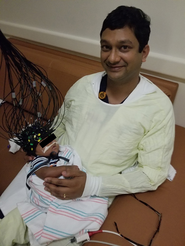
The precise imaging through CTOT, pronounced “see tot,” helps physicians accurately diagnose the severity of a baby’s brain injury and identify ideal treatment to optimize quality of life throughout childhood.
Disorders such as cerebral palsy, a birth-related stroke, and epilepsy affect an infant’s brain development. In the U.S., around 10,000 babies are born each year with cerebral palsy, and about 470,000 children have epilepsy, a seizure disorder. Currently there is no way to accurately capture brain activity to quantify the severity of these conditions without putting the infants to sleep for magnetic resonance imaging (MRI) or positron emission tomography (PET), since both require being still.
“Imaging helps us understand what parts of the brain are not functioning normally, which is critical for locating a seizure focus in epilepsy and understanding brain dysfunction after stroke, among other neurological diseases,” said Manish N. Shah, MD, assistant professor in the Division of Pediatric Neurosurgery with McGovern Medical School at UTHealth and pediatric neurosurgeon with UT Physicians and UTHealth Neurosciences. “MRI and PET scanning are both expensive, housed in other areas of the hospital, and require anesthesia for small children, so we had to find a new way to get the kind of functional brain mapping we needed.” Shah is also the director of pediatric spasticity and epilepsy surgery at Children’s Memorial Hermann Hospital.
To create the cap that babies can wear bedside while in the caregiver’s arms, Shah teamed up with Banghe Zhu, PhD, assistant professor with the Center for Molecular Imaging and Eva Sevick, PhD, professor and director of the center at the Brown Foundation Institute of Molecular Medicine for the Prevention of Human Diseases at McGovern Medical School. The first-generation device captures the imaging in minutes, and subsequent designs should allow for imaging in seconds.
“The cap is put on the child and harmless near-infrared light is passed through from one side and collected on the other side of the cap. Using a sensitive detector system, we can use the collected light to rebuild a 3D high-resolution picture of brain activity,” Shah said. “When we know what part of the brain is not functioning, we can either remove it, like in epilepsy; or stimulate it, like in epilepsy, cerebral palsy, Parkinson’s disease, or depression, etc. Better diagnosis yields more precise treatment and an improved outcome for the patient.”
Shah took the idea of an imaging cap to Sevick and Zhu at the Center for Molecular Imaging.
“We needed to find a way to detect very weakly transmitted near-infrared light and avoid interference. To overcome this, we adapted a ‘night-vision goggle’ technology,” said Zhu, the lead biomedical optical engineer on the project.
The laser light is harmless for the baby – the light is dimmer than the laser diodes used in a grocery store scanner, said Sevick, who oversaw the project that incorporated some of her already-developed technology.
“The military uses night goggles to detect the near-infrared heat signatures. Zhu is using the same night goggle technology to detect dim, near-infrared light transmitted across the brain of newborns. From the collected light, Zhu then uses a mathematical algorithm to determine a map of light-absorbing hemoglobin levels, which can provide clinical information of brain injury,” said Sevick, the Nancy and Rich Kinder Distinguished Chair in Cardiovascular Disease Research at McGovern Medical School. “No one has ever before been able to create an optical device that is sensitive enough for quickly imaging across the brain.”
It took about three years of developing prototypes, trial and error, and close communication between the physician and scientists for the cap to achieve precise whole-brain imaging in a clinical setting.
“I had never worked on a project from bench to bedside, so this is really a unique project for me,” Zhu said.
“It’s really rare for ideas to go from the bench to the bedside so quickly and successfully as our team executed,” Sevick said. “Success takes a special clinician to be patient with the ‘quirks’ of the engineers while maintaining the primary focus on clinical care. Our engineering team also listened carefully to the clinicians and worked in partnership with them to yield this exciting outcome.”
Because the device can be worn while awake and active, it opens the door for researchers and physicians to revolutionize diagnosis and treatment for movement disorders like cerebral palsy.
“The next step is to make an even higher resolution device and continue collecting data from children with strokes and epilepsy to better understand the diseases,” Shah said.
The project was funded by grants from the Memorial Hermann Foundation and a Men of Distinction award to Shah for excellence in community achievement.
Original post https://alertarticles.info

