The National Institutes of Mental Health awarded Yongsoo Kim, assistant professor of neural and behavioral sciences, $3.8 million to lead the project. His collaborators include Lydia Ng from the Allen Institute for Brain Science, James Gee from the University of Pennsylvania, Jiangyang Zhang from New York University and Luis Puelles from the University of Murcia in Spain.
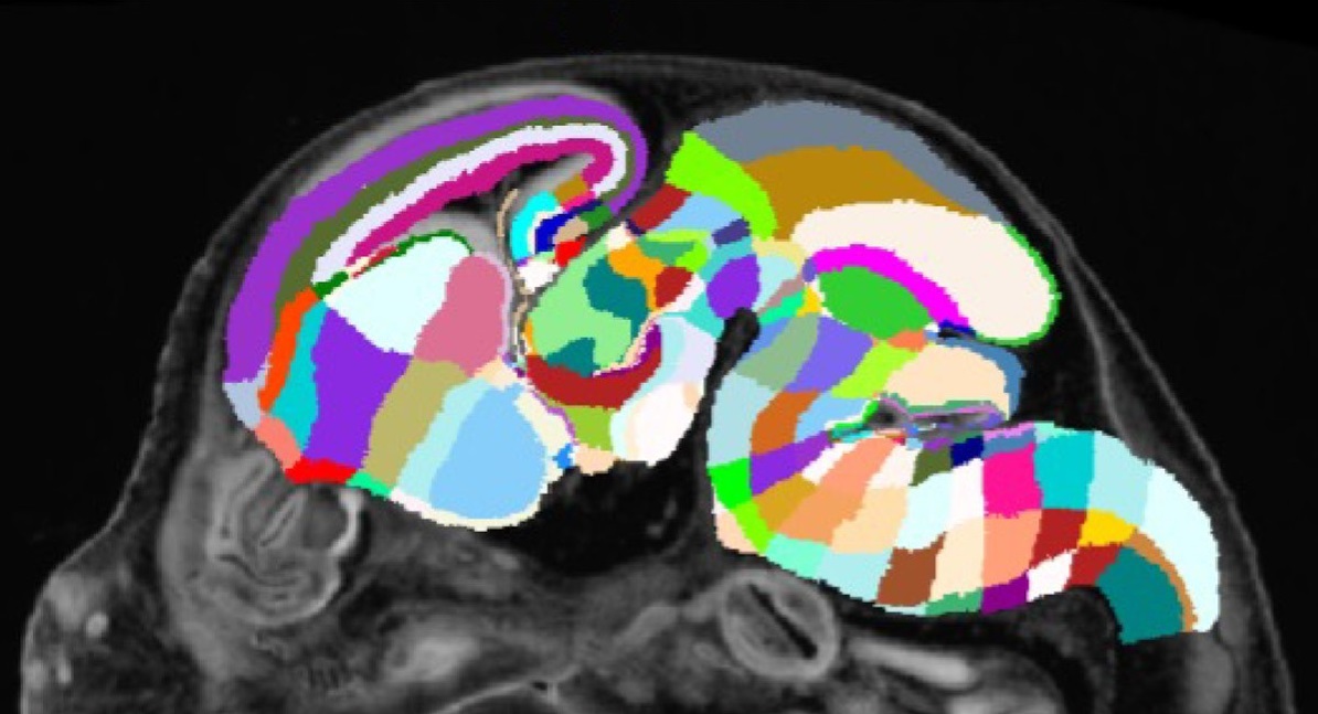
The research team will use computational tools and high-resolution 3D imaging to build a map of mouse brains across seven key developmental time points. Another goal of the project is to capture the location and quantity of key developmental neurons and other cell types across brain regions as the brain develops across time.
“There are many different types of cells that develop and connect across the brain during development and maturation,” Kim said. “By understanding the quantity and location of these cells in different brain regions, we can develop a baseline model against which we can measure changes that may occur in brain disorders.”
The project is funded through the National Institutes of Health’s Brain Research through Advancing Innovative Neurotechnologies (BRAIN) Initiative that aims to revolutionize understanding of the human brain by filling knowledge gaps of how the brain enables to the human body to record, process, use, store and retrieve information. The BRAIN initiative hopes to accelerate the development and application of innovative technologies that can show how individual cells and complex neural circuits interact in both time and space.
To establish the developmental atlases, the researchers will use special imaging and microscopy techniques to generate images of brain anatomy at seven developmental time points. They will then create 3D labels for key anatomy in those images and show how different regions change or connect across development and maturation. They will also map a special type of brain cell called GABAergic neurons, which serve as brakes for brain cell activity. Dysfunction of GABAergic neurons has been implicated in brain disorders and Kim said mapping the presence of these cells throughout development may help researchers understand how the brain makes decisions and processes external signals properly.
“If we can map the location and connections between GABAergic in neurotypical mouse models, we can compare them against models of schizophrenia and autism and try to identify the differences in brain development that may be causing these disorders to occur,” Kim said.
This project builds off of Kim’s work using high-resolution 3D imaging with computational tools to measure oxytocin receptor expression changes in developing mouse brains. He said that brain development tends to be similar across animal species and that developing these atlases for mice may help scientists better understand human brain development and disorders.
About Penn State College of Medicine
Located on the campus of Penn State Health Milton S. Hershey Medical Center in Hershey, Pa., Penn State College of Medicine boasts a portfolio of nearly $100 million in funded research. Projects range from development of artificial organs and advanced diagnostics to groundbreaking cancer treatments and understanding the fundamental causes of disease. Enrolling its first students in 1967, the College of Medicine has more than 1,700 students and trainees in medicine, nursing, the health professions and biomedical research on its two campuses.

