During the COVID-19 pandemic, Johns Hopkins Medicine Media Relations is focused on disseminating current, accurate and useful information to the public via the media. As part of that effort, we are distributing our “COVID-19 Tip Sheet: Story Ideas from Johns Hopkins” every other Tuesday starting July 28, 2020.
We also want you to continue having access to the latest Johns Hopkins Medicine research achievements and clinical advances, so we are issuing a second tip sheet covering topics not related to COVID-19 or the SARS-CoV-2 virus. Starting August 4, 2020, “Research News Tip Sheet: Story Ideas from Johns Hopkins Medicine” will alternate Tuesdays with the COVID-19 Tip Sheet.
Stories associated with journal publications provide a link to the paper. Interviews with the researchers featured may be arranged by contacting the media representatives listed.
PREVALENCE OF CHRONIC KIDNEY DISEASE AMONG MEXICAN AMERICANS HAS DOUBLED IN RECENT YEARS
A new study looking at nearly three decades of data from some 54,000 people has determined that the overall prevalence of chronic kidney disease (CKD) for several racial/ethnic and socioeconomic groups in the United States has stabilized in recent years, except Mexican Americans.
Results from the research, conducted by investigators from the U.S. Centers for Disease Control and Prevention (CDC) and three medical institutions — including Deidra Crews, M.D., associate professor of medicine at the Johns Hopkins University School of Medicine — were reported in the July 16, 2020, issue of JAMA Network Open.
Prevalence, as defined by the CDC, is “the proportion of persons in a population who have a particular disease or attribute at a specified point in time or over a specified period of time.” The study team looked at CKD prevalence among adults age 20 or older from 1988 to 1994 and from 1999 to 2016 for four racial/ethnic groups (non-Hispanic white, non-Hispanic Black, Mexican American and other), three educational levels (less than high school, high school and more than high school) and three income levels (low, middle and high).
“We found that overall prevalence of CKD, as well as the rates for all of the groups except one — Mexican Americans — stayed significantly the same from 2005 through 2016,” says Crews. “Among Mexican Americans during that period, the prevalence doubled, even when our analyses accounted for several potentially confounding variables, such as age and sex.”
Crews says the finding is consistent with recent studies showing a worsening overall health status for the Mexican American population compared with non-Hispanic whites.
Another revelation from the study, Crews adds, is that although CKD prevalence rates stabilized between 2005 and 2016 for most of the racial/ethnic groups and socioeconomic levels analyzed, significant disparities in who gets CKD still remain across racial/ethnic groups and socioeconomic levels.
“To narrow the gaps and move closer to health equity, stronger efforts are needed to effectively correct these persistent disparities in kidney health,” Crews says.
Data for the study came from the National Health and Nutrition Examination Survey (NHANES), a federal program to assess the health and nutritional status of adults and children in the United States over long periods of time. NHANES findings are used to determine the prevalence of major diseases and risk factors for those illnesses.
GENE THERAPY ALLEVIATES OBSTRUCTIVE SLEEP APNEA IN MICE
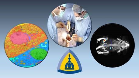
Floppy, untoned muscles in a person’s tongue and airway can block airflow and result in obstructive sleep apnea — a common disorder in which breathing repeatedly starts and stops during sleep, sometimes hundreds of times each night. Sleep apnea can cause daytime tiredness, increase the chance of accidents from falling asleep while driving, disrupt metabolism and if left untreated for years, lead to depression, high blood pressure, heart disease, stroke or death. Now, Johns Hopkins Medicine researchers have shown in mice that they can treat the condition and keep the airway open by using gene therapy to stimulate the nerve that contracts muscles in the tongue.
The researchers say their findings, published online on July 16, 2020, by the American Journal for Respiratory and Critical Care Medicine, ultimately may be translated into therapy for people at greatest risk of death from obstructive sleep apnea’s impact, such as those with conditions like stroke or severe atherosclerotic heart disease.
“The tongue muscles that are partly responsible for obstructive sleep apnea have a direct line via a nerve to the area of the brain that controls them,” says senior author Vsevolod Polotsky, M.D., Ph.D., director of sleep basic research and professor of medicine at the Johns Hopkins University School of Medicine. “Knowing this, we looked for a way to use this pathway in two directions: first, to travel to the control area with gene therapy to make it receptive to drug stimulation, and then once it’s been activated, send a signal back to the tongue to make the problem muscles contract.”
Obstructive sleep apnea affects 30% of adult men and 15% to 20% of women, says Polotsky. According to current medical practice, the gold standard for treating the condition is a continuous positive airway pressure (CPAP) machine that provides a steady flow of air to the user during sleep. “However,” he adds, “only 50% of people adhere to the treatment, so our team has been investigating alternative therapies.”
The tongue has eight pairs of muscles. One of these sets, the genioglossus, is stimulated by the hypoglossal nerve, which runs to it from the medulla, the lowest part of the brainstem. The medulla is connected to the spinal cord and controls involuntary functions, such as breathing and heartbeat. By implanting a pacemaker in the tongue, other researchers have shown that electrical pulses can make the hypoglossal nerve contract the genioglossus and prevent sleep apnea. The problem with this procedure, Polotsky says, is that it requires invasive surgery.
In their study, Polotsky and his team turned to a non-invasive method — an innocuous virus — to deliver laboratory-developed chemical receptors to the brains of obese mice. Once in place within the medulla, these receptors — known as DREADDs (Designer Receptors Exclusively Activated by Designer Drugs) — enable the brain cells in the medulla to accept a synthetic (“designer”) drug. The artificial DREADD receptors remain in the medulla for months after a single tongue injection.
After Polotsky and his colleagues injected the drug under the skin of the obese mice, it traveled up the hypoglossal nerve to the waiting DREADDs and stimulated the brain cells, which in turn activated the nerve to send a return signal to contract the tongue’s genioglossus muscles.
The researchers used magnetic resonance imaging on the sleeping mice to show that their airways opened after delivering the drug, but not in other mice injected with only saline. The researchers also measured six-fold higher electrical activity in the tongue muscles of the drug-treated mice, which indicated the tongue was contracting.
In upcoming mouse studies, the researchers plan to refine the virus delivery system to ensure it only gets DREADDs into the type of brain cells that control the hypoglossal nerve, rather than other nearby neurons. They also will look at the role that other tongue muscles may play in obstructive sleep apnea.
Based on these studies, human trials of DREADDs may follow.
“We believe that if this treatment works in humans, the drug to activate the receptors could be given orally every night and its effects will last about five to six hours,” says research associate Thomaz Fleury Curado, Ph.D., who led the study. “Therefore, by the time a person gets up, talking and eating won’t be a problem.”
NATIONAL STUDY SHOWS KIDNEY TRANSPLANTATION BETWEEN PEOPLE WITH HIV IS SAFE AND SUCCESSFUL
Kidney transplantation from deceased donors who had the human immunodeficiency virus (HIV) to people living with HIV and end-stage kidney disease is safe and consistently yields positive outcomes, according to researchers at Johns Hopkins Medicine and 13 other medical institutions. They say their finding could pave the way for more HIV-positive organs of all types — not just kidneys — being available for lifesaving transplants for people with HIV who need them.
The study was posted online on July 23, 2020, by the American Journal of Transplantation. It was performed under the authority of the HIV Organ Policy Equity (HOPE) Act, passed by Congress and signed into law in 2013. The act allows organ transplants from donors with HIV to recipients with HIV in approved U.S. research studies.
“This is an exciting culmination of 10 years of work: estimating the national impact of HIV-to-HIV transplants in 2011, helping write the HOPE Act in 2013, performing the first HIV-to-HIV transplants in the United States in 2016, and now collaborating across the country to demonstrate the safety of this procedure,” says Dorry Segev, M.D. Ph.D., professor of surgery and epidemiology at the Johns Hopkins University School of Medicine and co-leader of the HOPE in Action study team. “Fully successful implementation of the HOPE Act could mean hundreds of additional transplants and hundreds of additional lives saved each year.”
Between March 2016 and July 2019, the HOPE in Action team enrolled 75 adults with end-stage kidney disease and HIV whose virus was suppressed by anti-HIV therapy. Of the participants, 25 received kidneys from deceased donors with HIV and 50 received kidneys from deceased donors without HIV.
All participants survived transplantation, with median follow-up of 1.4 years for recipients of HIV-positive kidneys and 1.8 years for recipients of HIV-negative kidneys. At one year after transplantation, overall graft survival was excellent and comparable between HIV-positive kidneys (91%) and HIV-negative kidneys (92%). Additionally, there were no differences in hospitalizations due to infections, rates of serious adverse events or HIV-related complications, which were rare.
“People living with HIV face a higher risk of kidney failure than those without the virus, but their lack of access to suitable donor organs has previously meant many deaths while waiting for a transplant,” says Christine Durand, M.D., associate professor of medicine at the Johns Hopkins University School of Medicine and co-leader of the HOPE in Action study team. “So it’s a major step forward for our study to show that transplant outcomes using HIV-positive organs are comparable to HIV-negative ones.”
In a 2011 study, also published in the American Journal of Transplantation, a team led by Brian Boyarsky, M.D., a surgical research fellow and assistant resident at the Johns Hopkins University School of Medicine, estimated that 300 to 500 people with HIV could become deceased organ donors every year, potentially providing as many as 1,000 transplants annually for recipients living with the virus.
The new study provides solid evidence that the increase to the organ donor pool predicted nearly a decade ago is possible.
The work was primarily funded by the National Institute of Allergy and Infectious Diseases with additional support from the National Cancer Institute.
NEW JOHNS HOPKINS CENTER FOCUSES ON GENETIC EYE DISEASES
Often, one of the biggest challenges facing patients with a genetic condition is being able to diagnose it. For patients with one of the more than 350 known genetic eye disorders, the difficulty of making a diagnosis — and as a result, choosing the appropriate treatment — can be compounded because their eye disease may occur in isolation or as part of a broader, systemic genetic problem.
To address the needs of patients with genetic eye disorders, ophthalmologists at the Johns Hopkins Wilmer Eye Institute have established the Wilmer Genetic Eye Disease (GEDi) Center. The center offers coordination of care for patients with genetic eye disorders, along with dedicated genetic counseling and access to subspecialists in all fields of ophthalmology.
For some patients with a suspected systemic genetic condition, subtle eye findings may help confirm a diagnosis. Other patients may present with an isolated eye finding that is genetic but may not be readily recognized as genetic.
“If we identify a particular feature in the eyes that may suggest a systemic genetic problem, having the knowledge to send that person for work-up with other groups around the hospital is really important,” says Jefferson Doyle, Ph.D., M.B.B.Ch., M.H.S., assistant professor of ophthalmology and genetics at the Johns Hopkins University School of Medicine and co-leader, along with Mandeep Singh, M.D., Ph.D., assistant professor of ophthalmology at the medical school, of the GEDi team of specialists.
Having a diagnosis is important because it can tell the patient what is the likely course of a disease, whether family members are at risk, the chance that future offspring will be affected and what treatment options may be available. With a diagnosis in hand, patients also can learn if there are any clinical trials in which they could participate (or benefit from, if the trials produce new treatments).
Another plus for patients going to the GEDi center is access to an on-site genetic counselor. This is particularly important for patients who require genetic testing in order to participate in a clinical trial. Additionally, long wait times for counseling can affect the ability to treat a disease before it damages the eyes.
In recent years, significant advancements in the field of genetics have been made, and there are new opportunities for therapeutic intervention. Nowhere is this more apparent than in the field of ophthalmology, which pioneered some of the first gene therapies approved by the Food and Drug Administration (FDA). In 2017, for example, the FDA approved a gene therapy for Leber congenital amaurosis, a rare form of inherited vision loss. Doyle and Singh foresee many more gene therapies being developed — therapies that may greatly improve the quality of life for those affected by genetic eye diseases.
The GEDi center is already well-positioned to become a major center for genetic clinical trials. Current, completed and upcoming genetic clinical trials there include those for Stargardt disease, X-linked retinitis pigmentosa and juvenile glaucoma.
What really makes the GEDi center special, says Doyle, is the connection to Johns Hopkins and its broad spectrum of expertise, skills and facilities.
“Johns Hopkins is really an ideal place to provide this kind of care,” says Doyle. “You have this incredible wealth of knowledge and experience at the Wilmer Eye Institute, paired with one of the top departments of genetic medicine in the world. It provides us with such a wonderful opportunity to help patients with these challenging conditions.”
Original post https://alertarticles.info

