Winners will be highlighted on slides during the 2019 ASCB|EMBO Meeting in Washington, DC, Dec 7-11. The winners will also be recognized during a 7:15 pm, Tuesday, Dec. 10 session entitled, “GFP25: Lighting up Cell Biology,” with Nobel Laureate Martin Chalfie of Columbia University and Jennifer Lippincott-Schwartz of Janelia Research Campus. Both will offer an historical perspective and present their research using GFP.
“All the submissions to our contest were amazing and showcased the elegant and beautiful scientific images researchers are able to create using this simple protein,” said ASCB CEO Erika Shugart. “They were so impressive, both visually and scientifically, that I know it was a challenge to choose the very best ones. ASCB thanks everyone for participating in this first-time event. I also want to thank Marty and Jennifer for helping us close out the celebration of this important bioscience tool.”
There were 67 submissions to the contest. ASCB received entries from across the United States including Puerto Rico, as well as from Brazil, Chile, England, Finland, France, Israel, The Netherlands, and Poland. Congratulations to all the winners and thanks to everyone who submitted all of these beautiful images and videos.
Here are the winners in ASCB’s 2019 GFP Image and Video Contest. A link to the gallery of all the winners may be found at the top of the contest page here.
Images
First Place – Sumana Sundaramurthy, SUNY Upstate Medical University: The assembly of a worm rocket!
Three-way tie for Second Place –
Daniela Malide, National Institutes of Health: NIH3T3-co5FPs_ Mouse fibroblasts NIH-3T3 cells co-transduced with 5 fluorescent proteins.
Sumana Sundaramurthy, SUNY Upstate Medical University: Movement of screw-propelled worms!
Ivan Surovtsev, Yale University: MicroUniverse
Two-way tie for Third Place –
Mike Henne, UT Southwestern Medical Center: Adipocytes keep in touch with their fat stores
Joachim Goedhart, University of Amsterdam: Quintuple
People’s Choice
Bruno da Costa Rodrigues, Federal University of Rio de Janeiro: F-actin structure of a salivary gland from the kissing bug insect
Honorable Mentions
Page Baluch, Arizona State University: GFP Lifeact Mouse Uterus
Daniela Malide, National Institutes of Health: Ear-mouse myosinIIA-EGFP
Andrew Moore, Howard Hughes Medical Institute: Zebrafish F-actin
Bruno da Costa Rodrigues, Federal University of Rio de Janeiro: F-actin structure of a salivary gland from the kissing bug insect
Jennifer Heppert, Duke University: Not just “Green”
Videos
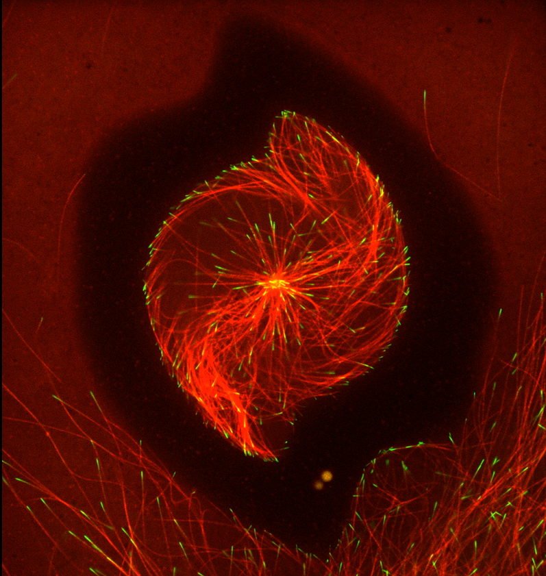
First place – Jesse Gatlin, University of Wyoming: Cytoskeletal Storm
Second place – Giulia Zarfati, Weizmann Institute of Science: Myoblast fusion
Third place – Jeffrey van Haren, UCSF (currently Erasmus MC): Microtubule and actin cytoskeleton in a human epithelial cell
People’s Choice
Giulia Zarfati, Weizmann Institute of Science: Myoblast fusion
Honorable Mentions
Thomas Di Mattia, Institute of Genetics and Molecular and Cellular Biology – Illkirch, France: Connected organelles in motion
Matthew Pittman, Johns Hopkins University: Focal Adhesion Fireworks
Pablo Saez, Institut Curie – IPGG (MOTILE team): The chemokine call
Jules Riley, Syracuse University: A Stressful Phase
Zhirong Wang, Georgetown University: Spontaneous Auditory Rehearsal
Original post https://alertarticles.info

